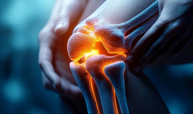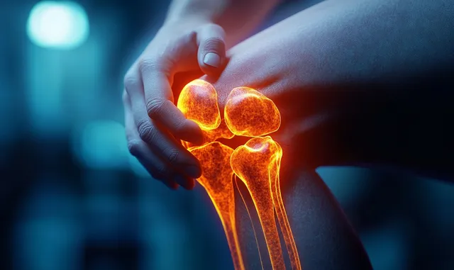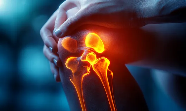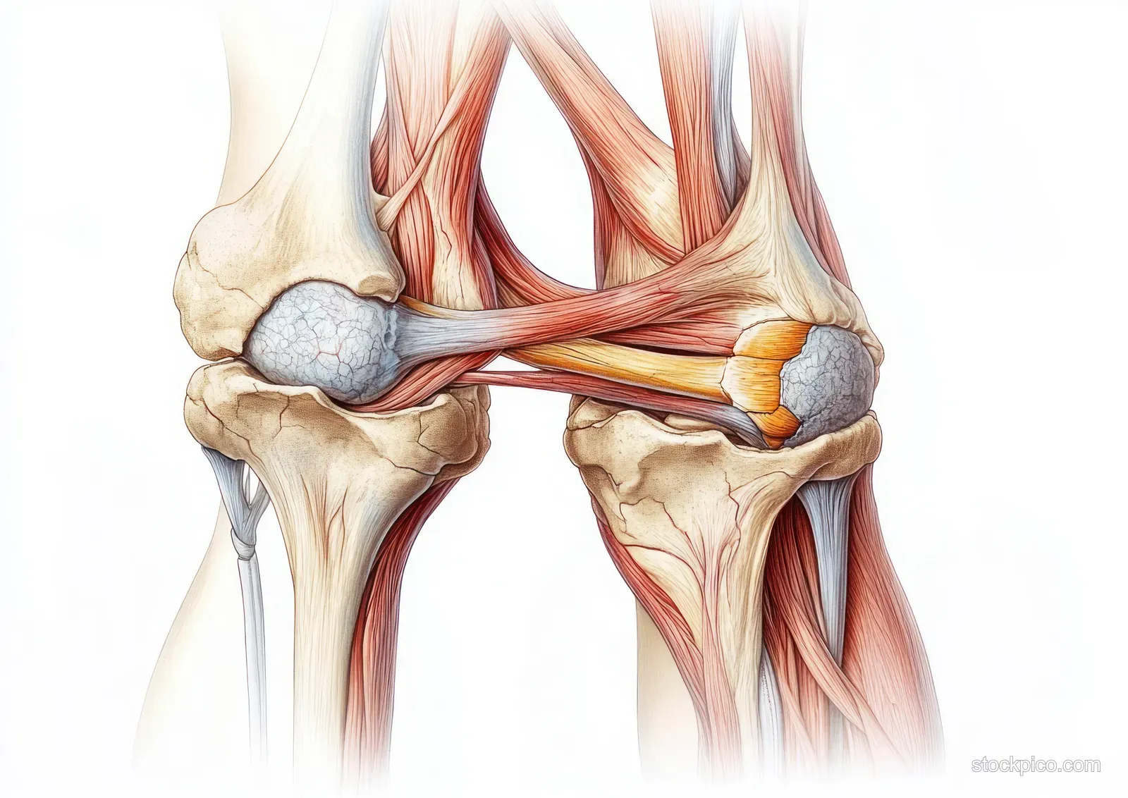
Cross-section anatomical knee illustration showing femur, patella, meniscus, and tendons
An educational medical diagram of the human knee joint in cross-section, highlighting muscles, ligaments, cartilage, and the meniscus around the patella and femur.
Related Editorial Assets