3d Anatomical Illustration images

Explore stock images tagged 3d anatomical illustration
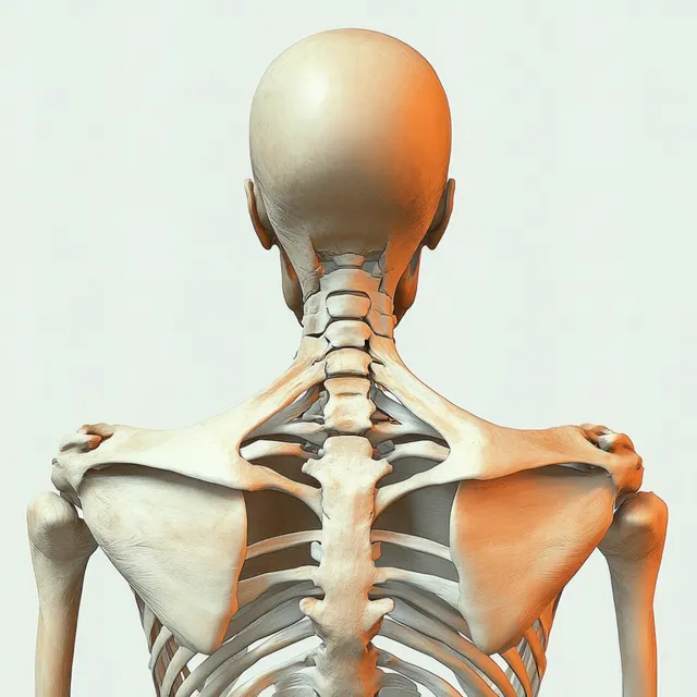
Rear view 3D anatomical illustration of a human skeleton showing spine, scapulae, and shoulders.
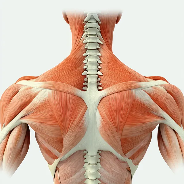
Back view 3D render of shoulder muscles over scapula and clavicle with ribs and spine partially visible.
3D knee joint anatomy with femur and tibia, highlighting an inflamed pain area using a bright orange glow.
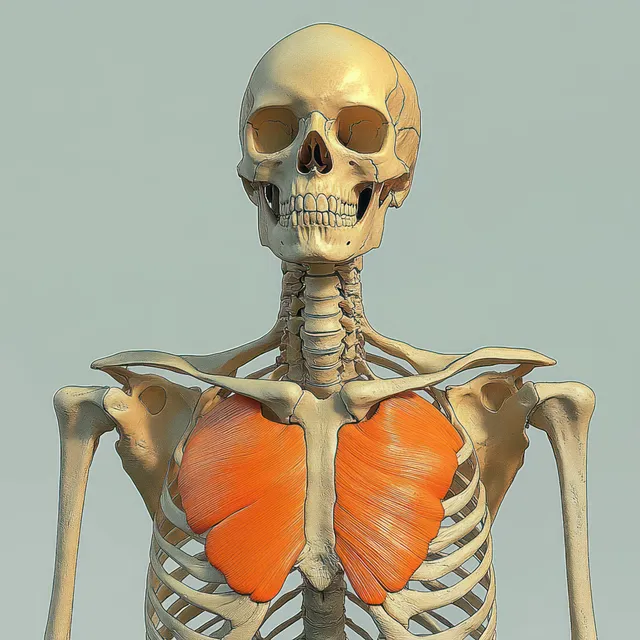
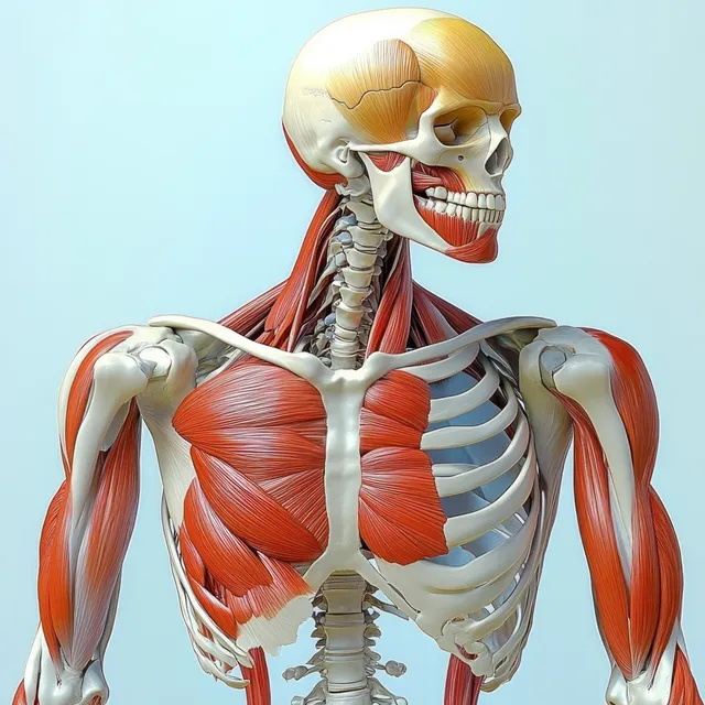
A human skeleton facing forward with orange-highlighted pectoral muscles across the ribcage.
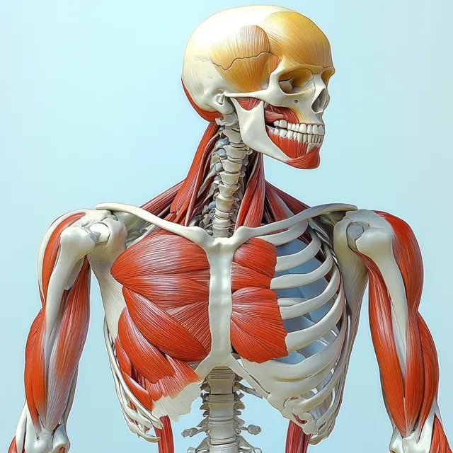
Front view 3D anatomy model showing human skeleton with red biceps brachii muscles highlighted.
3D anatomical illustration of a human skeleton with biceps brachii muscles highlighted on the upper arms.

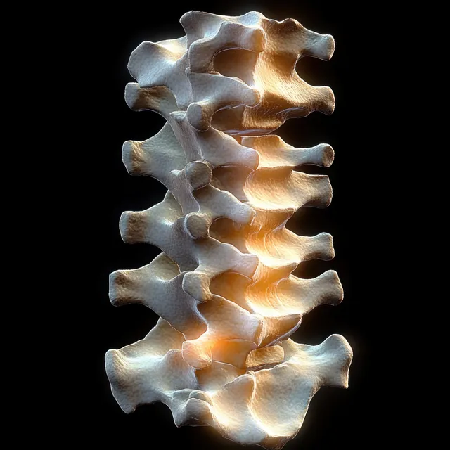
3D anatomical spine model with vertebrae and posterior longitudinal ligament isolated on a black background.
3D anatomical illustration of a human skeleton with biceps brachii muscles highlighted on the upper arms.
3D render of a human knee joint with a glowing red inflamed patella area on blue bones.
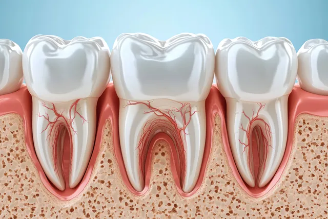
Cross section of teeth showing healthy gums beside inflamed recession and exposed roots.
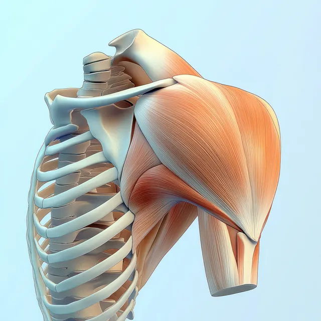
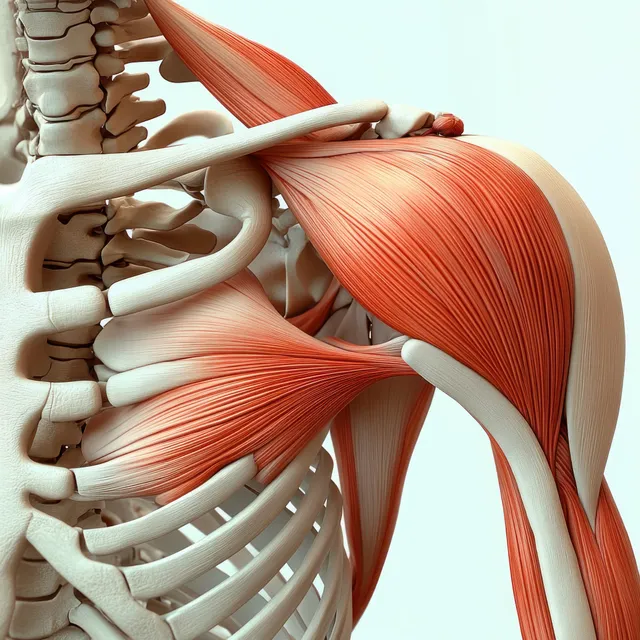
3D anatomical view of the deltoid muscle on the shoulder with clavicle and scapula visible behind.
Anterior shoulder anatomy model showing highlighted deltoid muscle with visible clavicle, scapula, and rib cage.
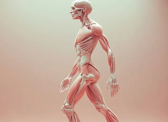
3D human figure anatomy view with transparent muscles and skeleton, showing labeled blood vessels on a soft background.
3D model of a fatty human liver with highlighted vessels floating over a blurred blue medical background.
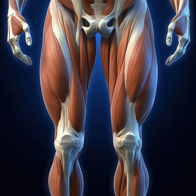
Male leg anatomy illustration highlighting quadriceps muscles with visible femur and tendons under blue dramatic lighting.
3D anatomical view of a glowing heart and vascular system inside the chest cavity.
Posterior upper-back anatomy illustration with visible spine and shoulder muscles including rhomboid major and trapezius.
A hyper-realistic 3D human brain model with detailed folds isolated on a white background.
3D ankle joint X-ray illustration with red inflammation highlights on a dark blue medical background.
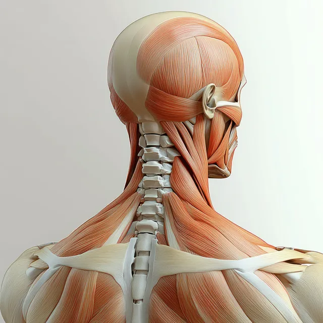
Rear view anatomical 3D illustration of the neck and trapezius muscles over the vertebrae.
Transparent blue torso with an orange glowing heart and branching blood vessels visible inside the chest.
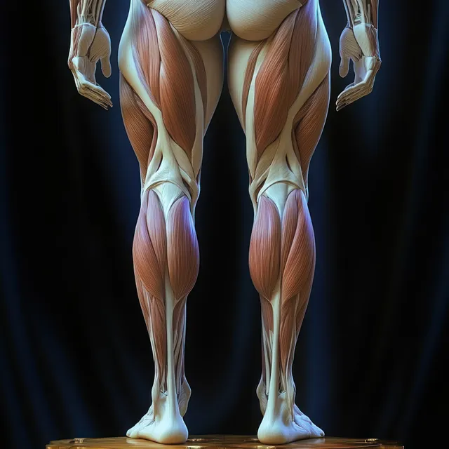
Female lower-body anatomical illustration displaying hip, thigh, and knee muscles with dark lighting and visible tendon lines.

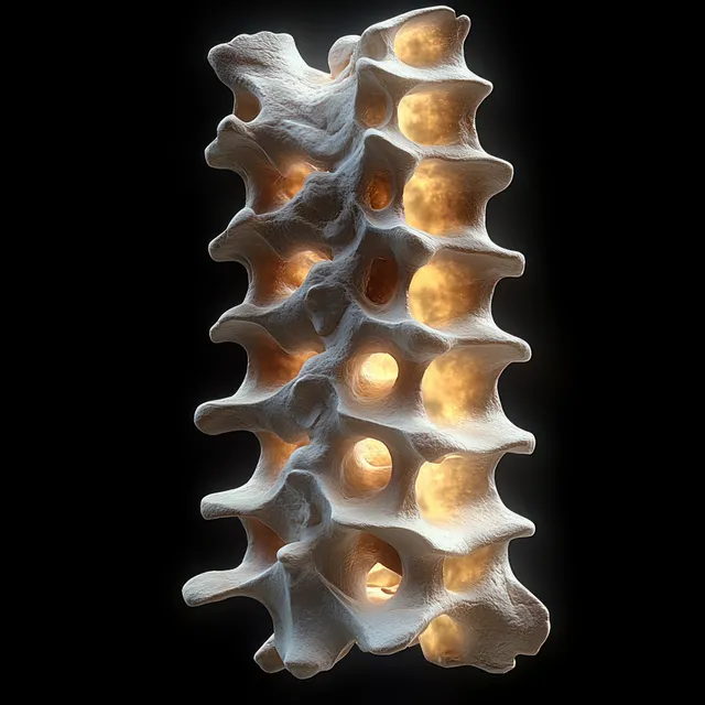
3D vertebral column model with illuminated spinal discs isolated on a dark background.