Anatomy Diagram images

Explore stock images tagged anatomy diagram

Side profile anatomical diagram showing labeled human brain and spinal cord with visible internal regions.
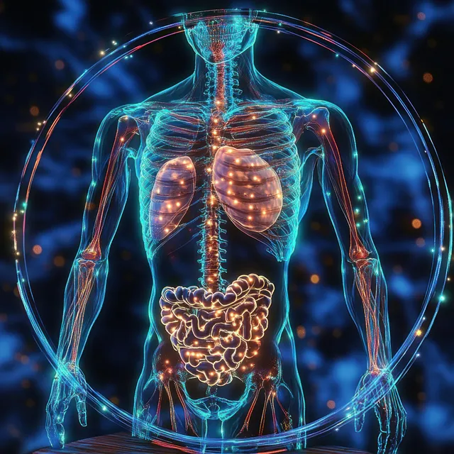
Bioluminescent skeletal ribs and glowing organs inside a circular frame surround a human torso illustration.
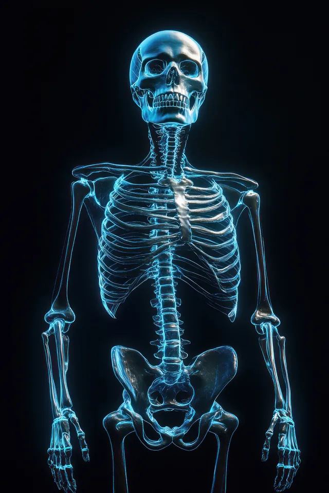
Neon outlined human skeleton render lit by a spotlight on a black background.
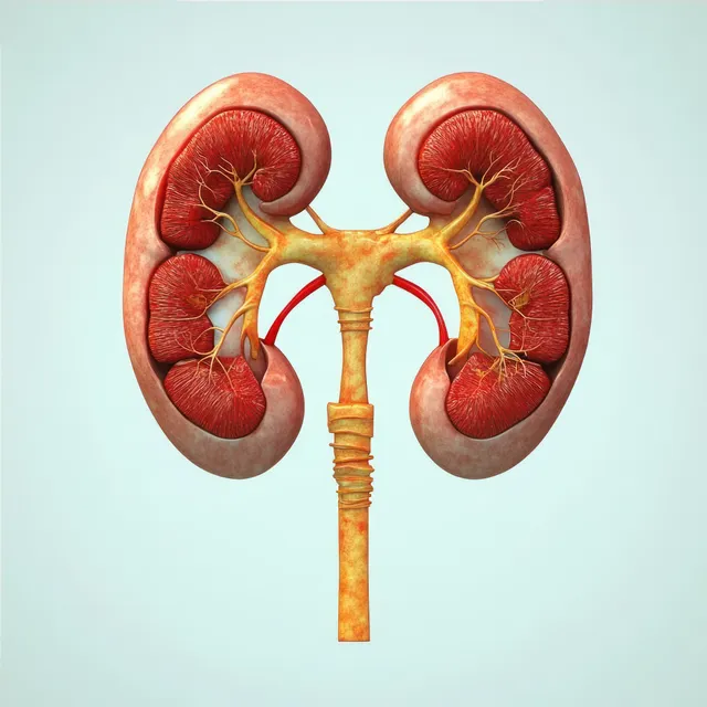
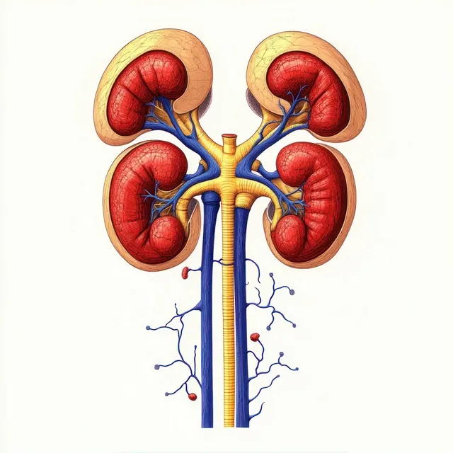
An anatomical kidney diagram displays renal arteries and veins with ureters on a light background.
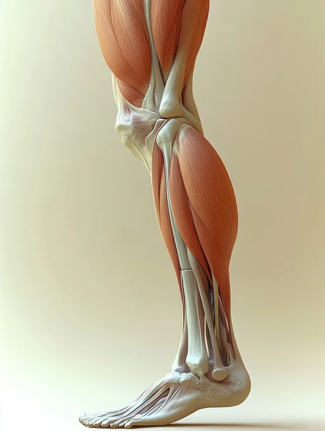
Lateral view anatomical illustration of the hamstring muscles and knee joint with highlighted thigh tendons.
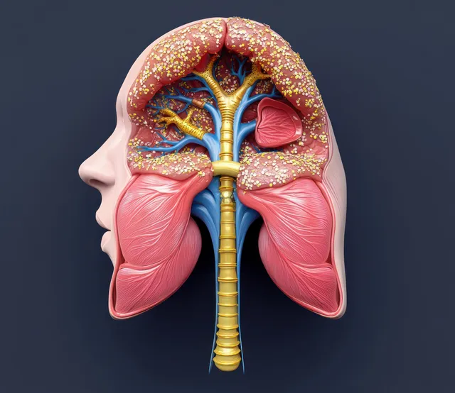
Cross-sectional anatomical illustration shows the human throat and nasal passages leading into the trachea and bronchi.
Four kidneys diagram showing ureters leading downward with connected blue blood vessels and internal renal regions.
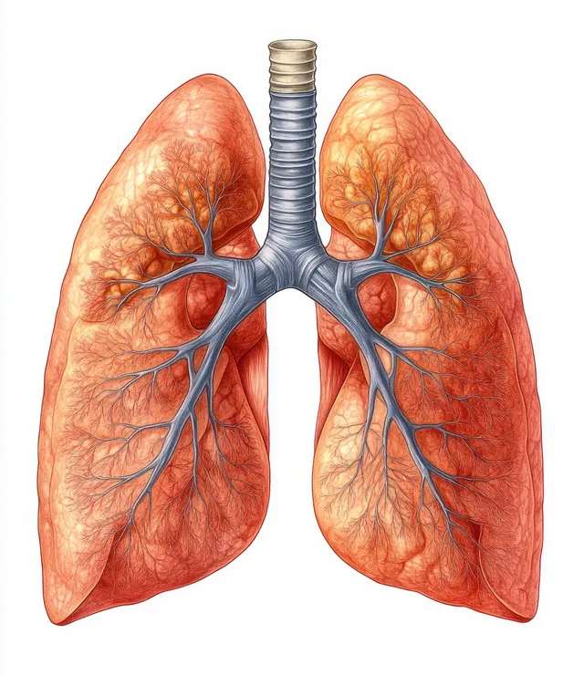
An anatomical illustration of human lungs with bronchi and visible vessels on a white background.
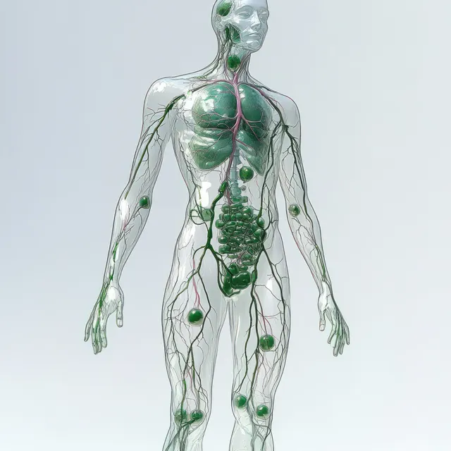
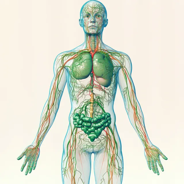
Transparent female figure with glowing green lymphatic vessels over the chest and abdomen against a light background.
Transparent female torso illustration displaying colored lymphatic vessels and lymph nodes across the chest, abdomen, and arms.
3D spine illustration with glowing orange pain areas along the vertebrae over a dark blue medical background.
Labeled shoulder joint anatomy illustration showing clavicle, scapula, humerus, and ligaments on a white background.
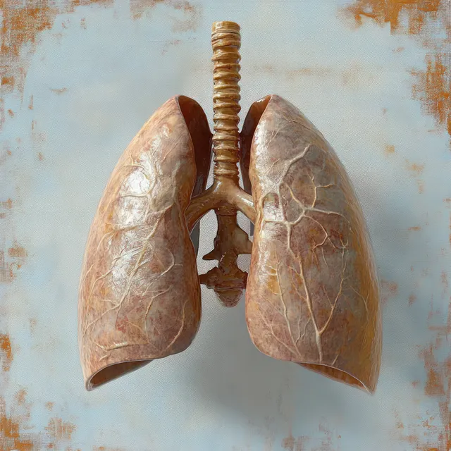
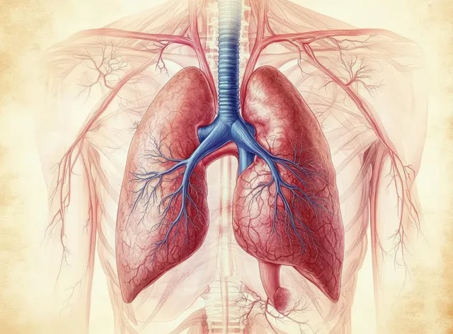
Detailed anatomical model of human lungs with visible trachea and bronchi on a pale blue textured background.
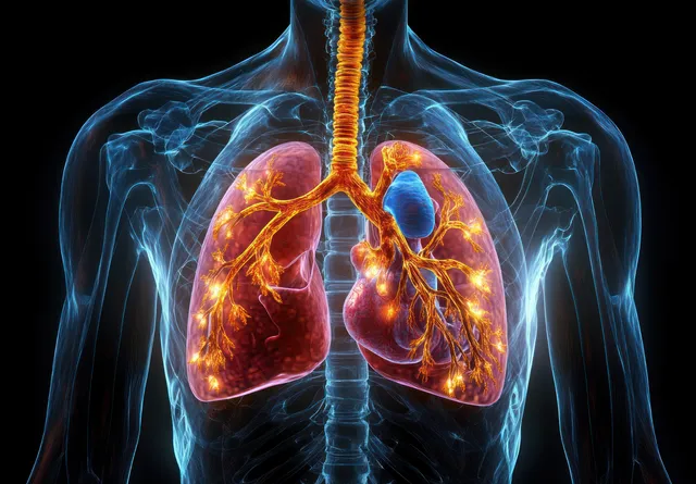
Translucent torso illustration shows glowing heart and lungs with bronchi and branching blood vessels on black.
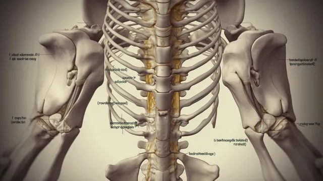
Labeled anatomical illustration of the human pelvis and spine shown in a symmetrical posterior view.
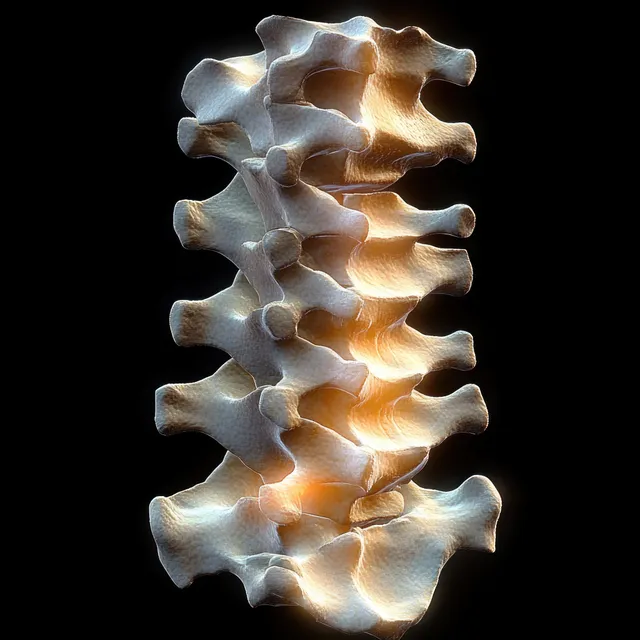
3D anatomical spine model with vertebrae and posterior longitudinal ligament isolated on a black background.
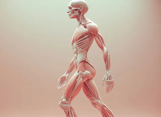
3D human figure anatomy view with transparent muscles and skeleton, showing labeled blood vessels on a soft background.
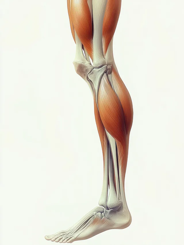
Side-view anatomical illustration of hamstring muscles on a human leg highlighting semitendinosus and biceps femoris longus.
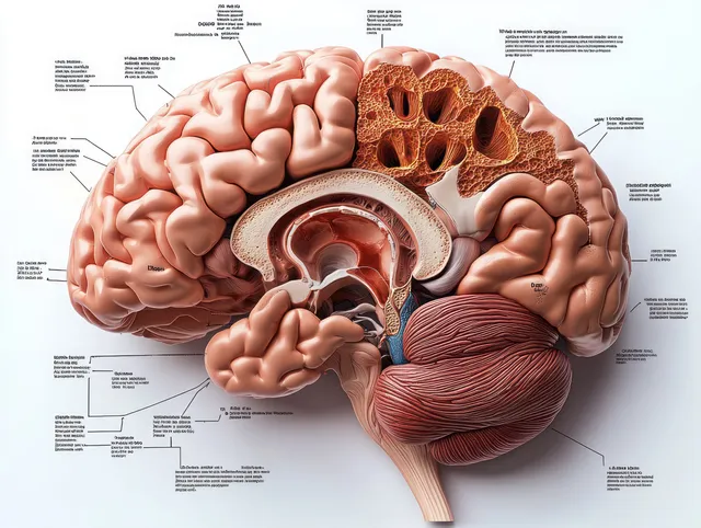
Labeled sagittal brain diagram showing frontal lobe and cerebellum in detailed anatomical section.
Cutaway stomach and esophagus illustration showing acid reflux and inflamed esophageal tissue near the junction.
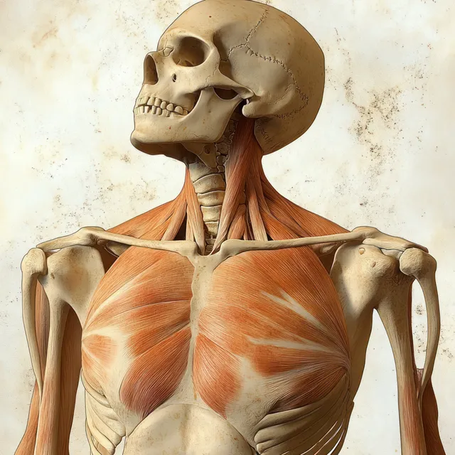
Skeleton model shown from the front with highlighted pectoralis major muscles and labeled chest anatomy.
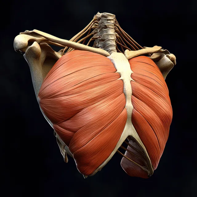
Red pectoral muscles are highlighted on an anterior skeletal torso against a dark background.
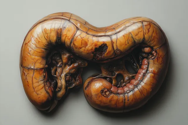
A surreal glowing stomach-like organ with visible internal folds in vivid amber and red tones.
Medical illustration of human lungs, heart, and branching bronchial tree with blue blood vessels inside the ribcage.
Anatomy Diagram images | StockPico