Anatomy Study images

Explore stock images tagged anatomy study

Transparent kidney anatomical model with visible kidney stones on a clear stand beside books, plants, and office supplies.
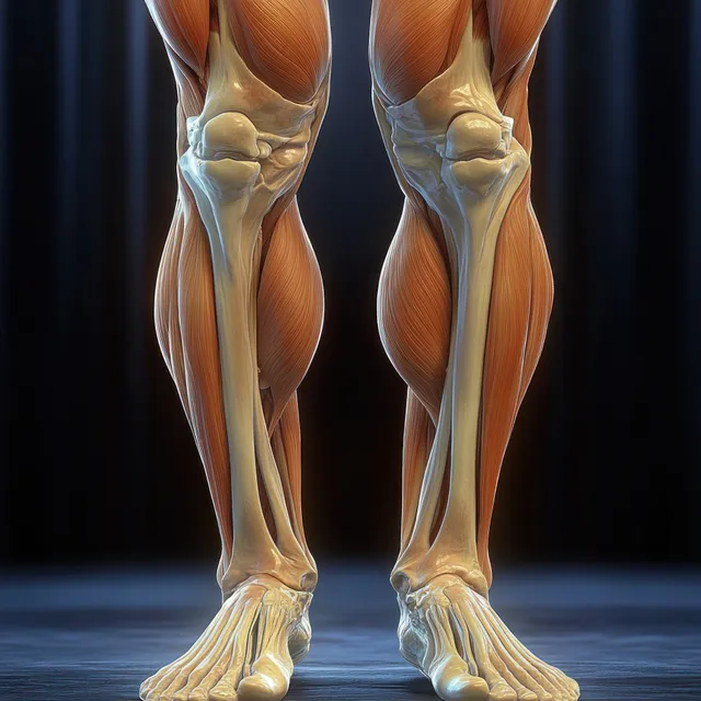
Anterior view 3D anatomical legs highlighting quadriceps muscles and knee joint with visible bone contours.
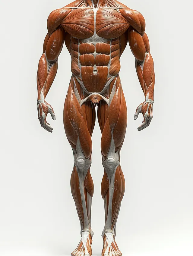
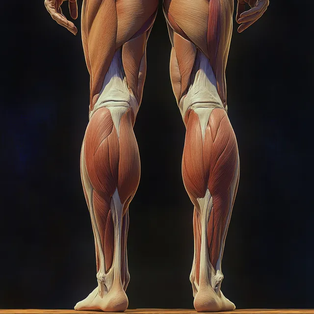
Standing male figure showing detailed leg muscle groups, tendons, knee, ankle, and foot contours on a plain background.
Yellowed human skull isolated on a white background with visible teeth, eye sockets, and cranial details.
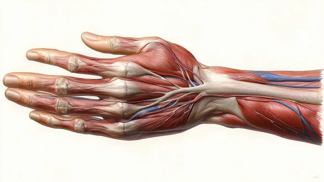
Anatomical illustration of a female hand ventral view with visible muscles, tendons, arteries, and veins.
A front view light brown human skull with prominent canine teeth against a plain white background.
Gray alien sculpture with long fingers and outstretched hands, illuminated in dramatic light against a dark background.
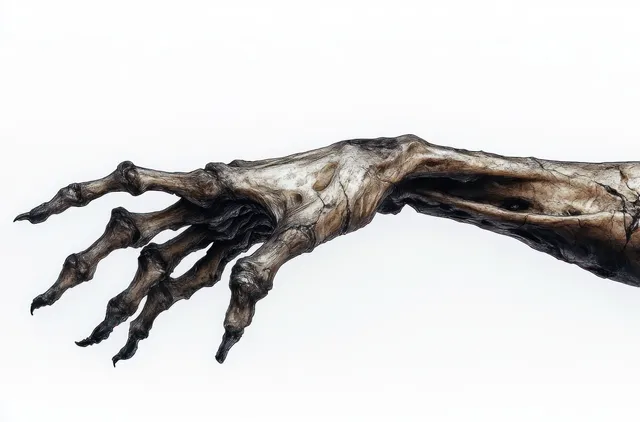
A twisted skeletal hand with elongated fingers stretches across a plain white background, showing detailed weathered bone texture.
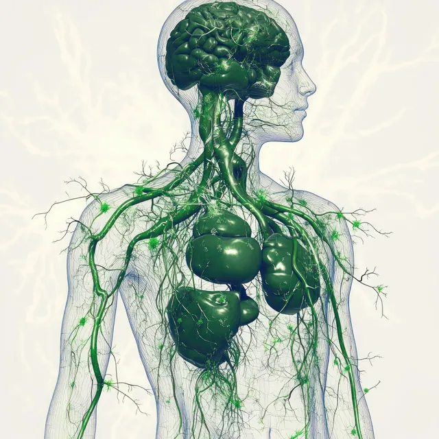
Green lymphatic vessels branch across a semi-transparent torso showing chest and abdomen organs.
Medical illustration of a human skeleton showing glowing lumbar spine area for lower back pain.
Human skeletons in dynamic floor poses are illuminated with dramatic chiaroscuro lighting against a dark textured background.
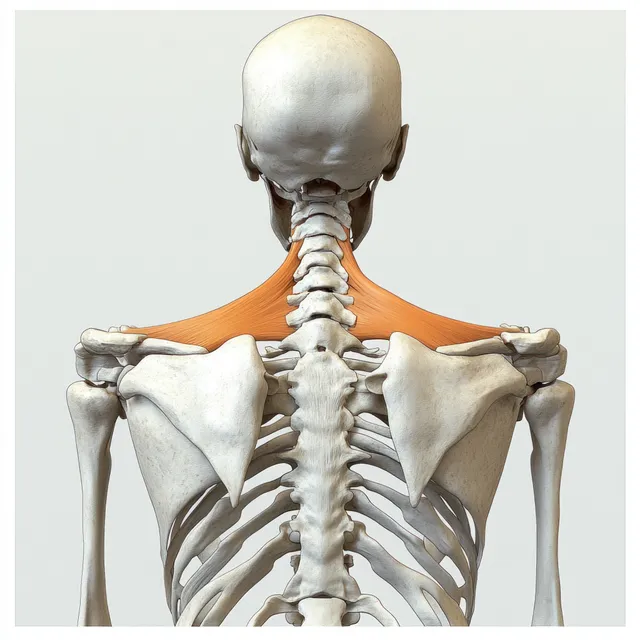
Rear-view human skeleton illustration highlighting the spine and shoulder blades under soft studio light.
A hand holds a pen pointing at the rib cage and spine on a detailed anatomical model under warm light.
Close-up of a human skull with cracked bone and visible teeth against a dark background.
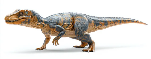
Baryonyx theropod dinosaur walking in side profile with detailed scales and teeth on a white background.
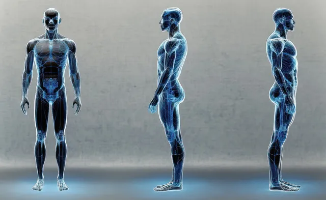
Three transparent male figures show front and side musculoskeletal anatomy on a textured gray background.
Glowing cyst-like shapes overlay ovarian and uterus anatomy in a pelvic medical illustration.
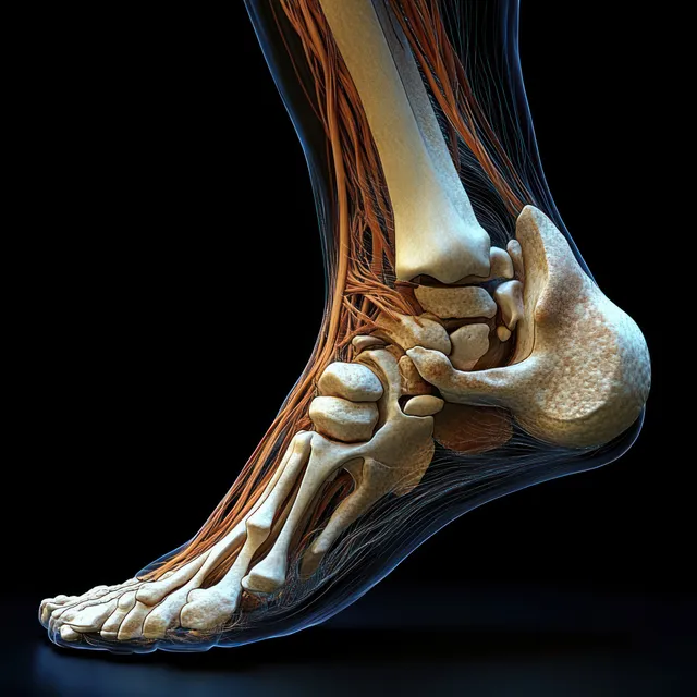
Medial x-ray style view of a human foot showing heel bones, ankle joints, and glowing tendons on a dark background.
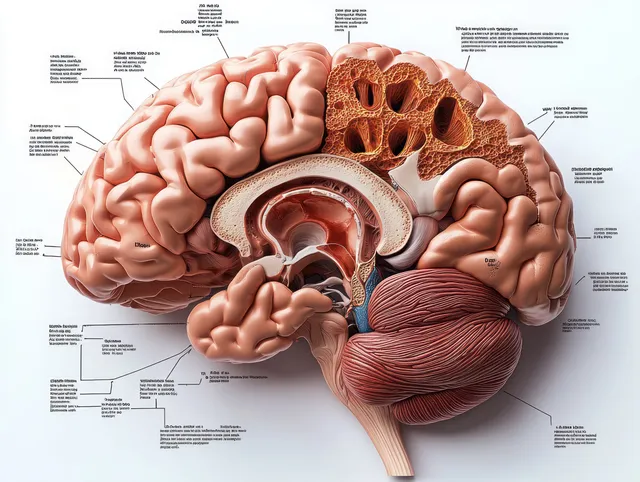
Labeled sagittal brain diagram showing frontal lobe and cerebellum in detailed anatomical section.
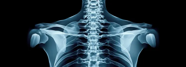
X-ray image shows upper back thoracic spine, ribs, and shoulder blades against a black background.
Posterior view of a stylized female leg with highlighted hamstrings and hip muscles over dark background.
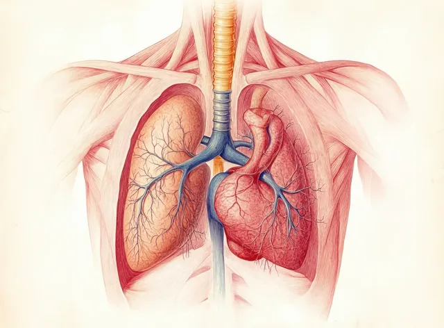
Illustrated human torso showing lungs and heart inside a rib cage with branching bronchial and blood vessels.
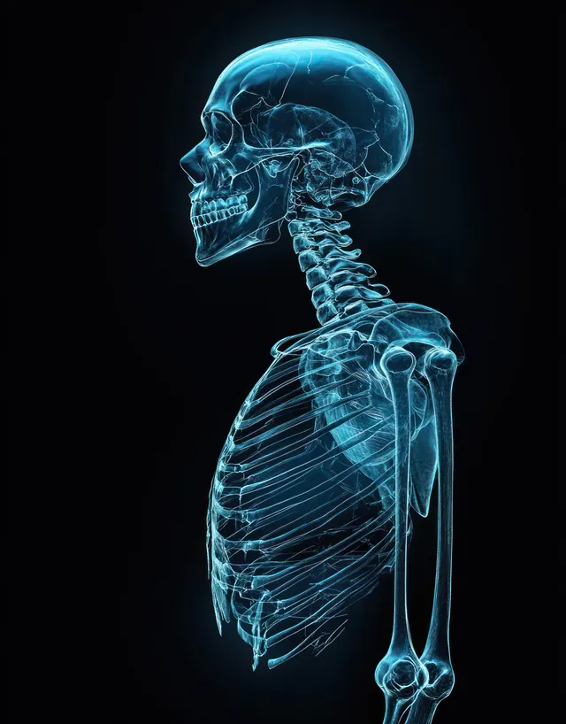
Glow-lit blue male skeleton in three-quarter view with skull, spine, ribs, and joints on a dark background.
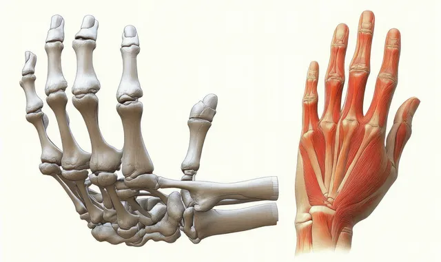
An anatomical illustration shows a human hand with bones, muscles, and tendons in dorsal view.