Anterior View images

Explore stock images tagged anterior view
Skeleton model shown from the front with highlighted pectoralis major muscles and labeled chest anatomy.
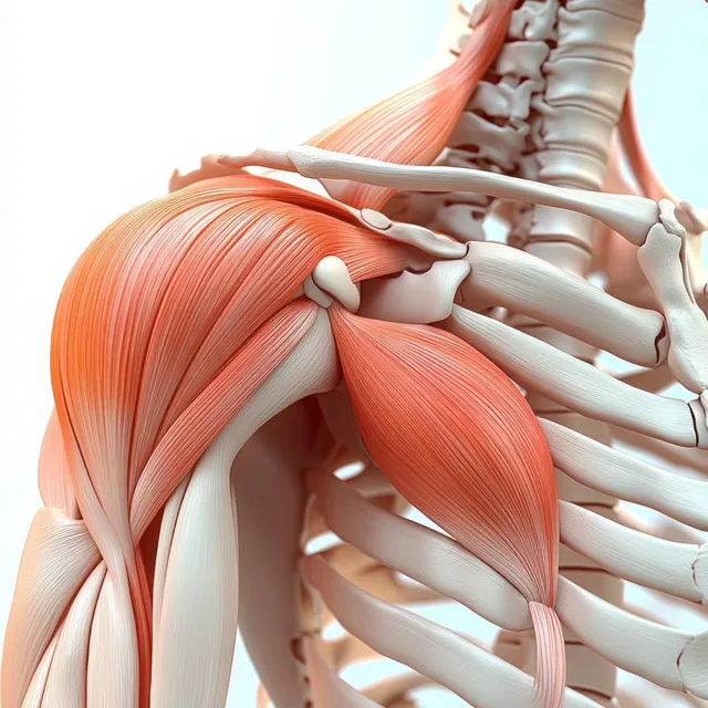
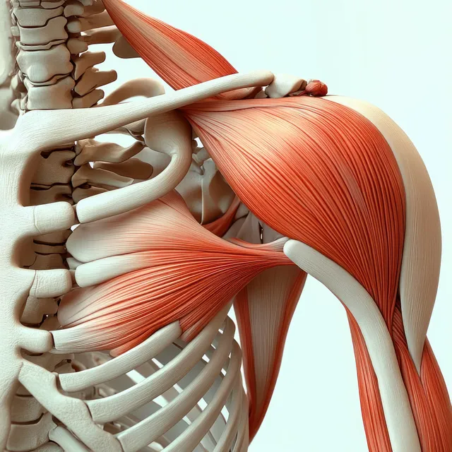
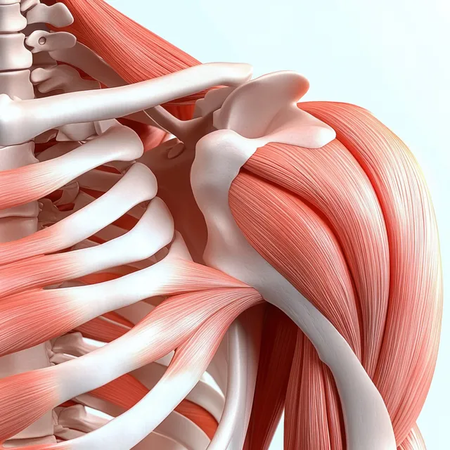
3D anatomical view of the deltoid muscle on the shoulder with clavicle and scapula visible behind.
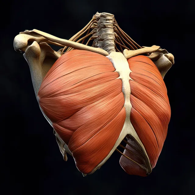
Red pectoral muscles are highlighted on an anterior skeletal torso against a dark background.
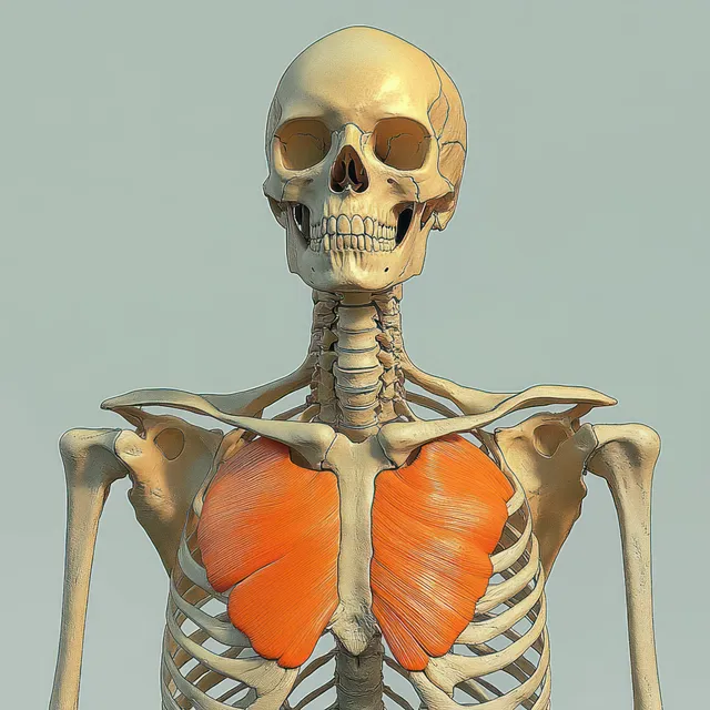
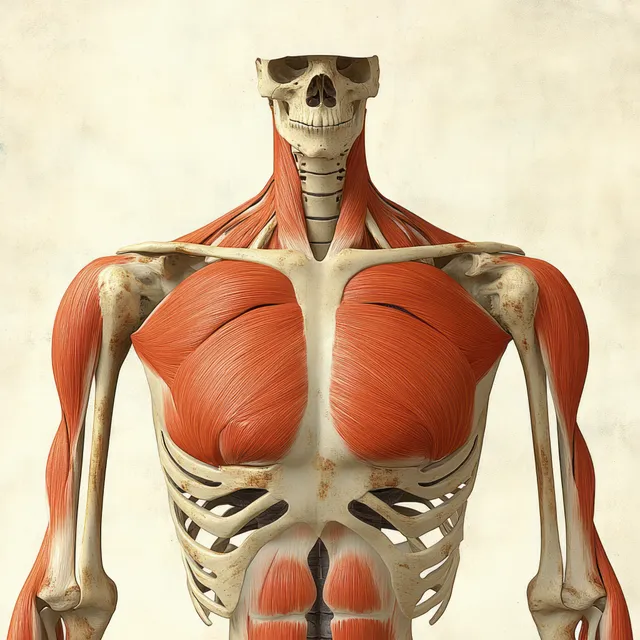
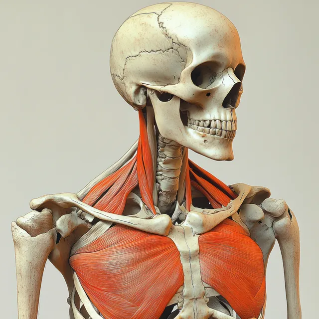
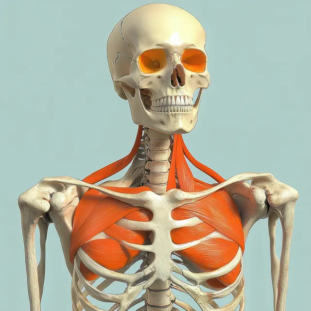
A human skeleton facing forward with orange-highlighted pectoral muscles across the ribcage.
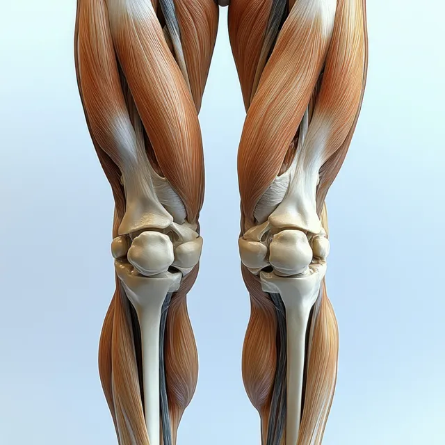
Anterior view 3D anatomical legs highlighting quadriceps muscles and knee joint with visible bone contours.
Anterior shoulder anatomy model showing highlighted deltoid muscle with visible clavicle, scapula, and rib cage.
Red pectoralis major muscles are shown on an anterior human skeleton with labeled ribs, clavicle, and shoulder bones.
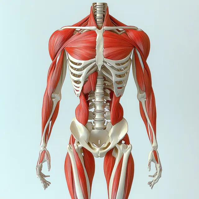
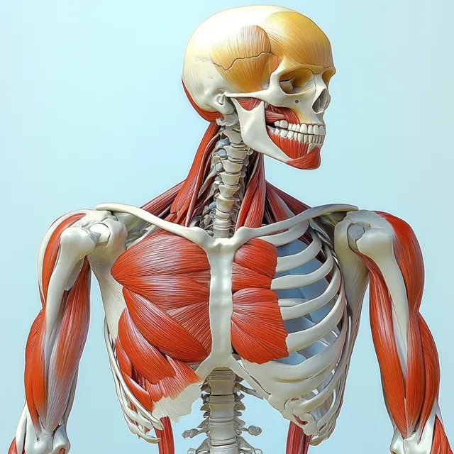
Anterior 3D illustration of the human skeleton with red biceps muscles, ribs, and shoulders on a light background.
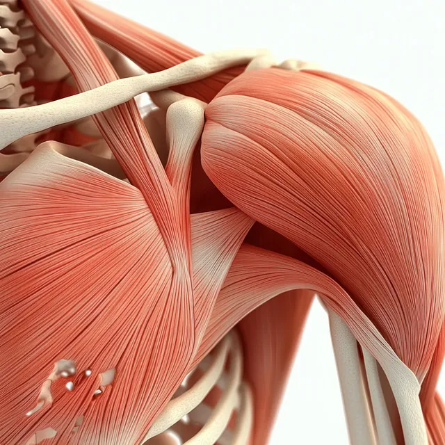
Anterior shoulder anatomy illustration highlighting the deltoid muscle, tendons, and surrounding bones.
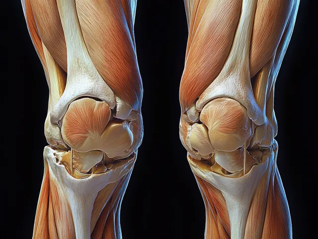
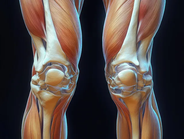
3D render of anterior knee anatomy showing femur, patella, meniscus, cartilage, and surrounding ligaments on a dark background.
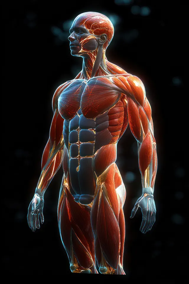
A backlit 3D figure shows glowing anterior muscles of the human torso and arms against a dark background.
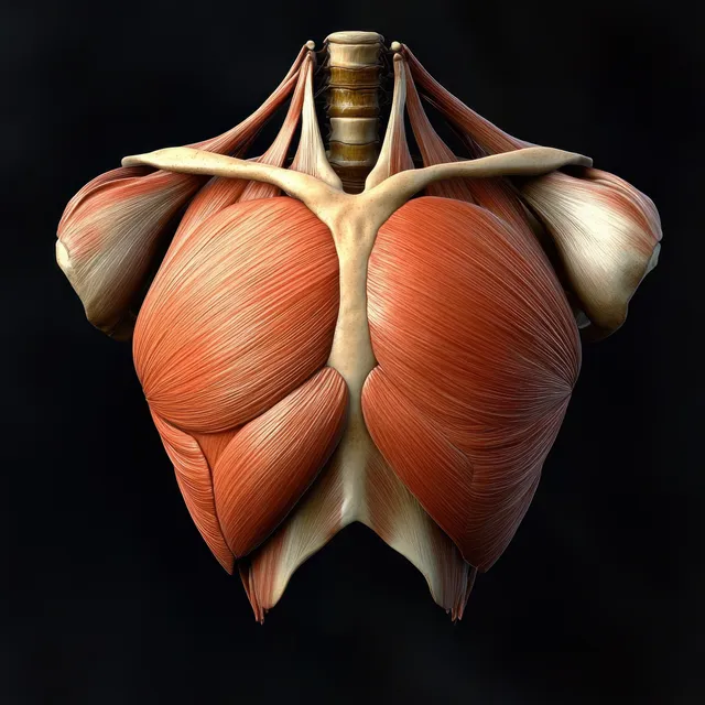
Anterior anatomical illustration of the pectoralis major muscle with ribs, clavicle, and humerus on a black background.
Color anatomical illustration highlights leg and arm muscles, tendons, nerves, and joints floating on a dark background.
Anterior view anatomical illustration showing the deltoid muscle with clavicle and scapula bones and surrounding tissue layers.
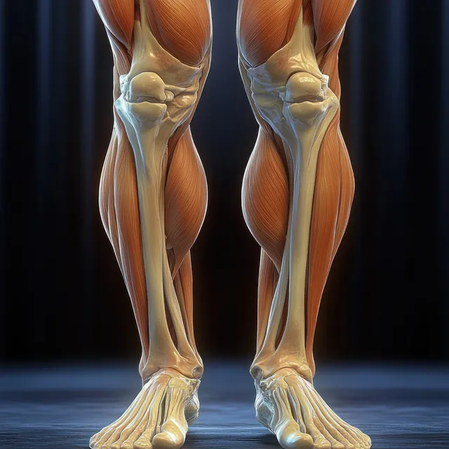
Anatomical illustration of both knees and thighs with visible quadriceps muscles, bones, and knee joint alignment against a plain background.
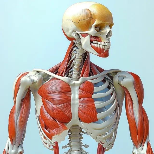
A front view anatomical illustration of a human skeleton with orange-highlighted muscles across the chest and neck.
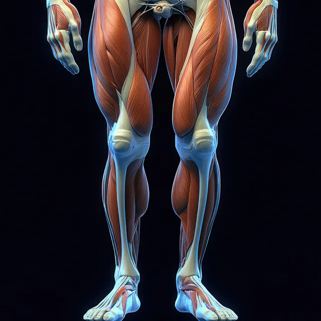
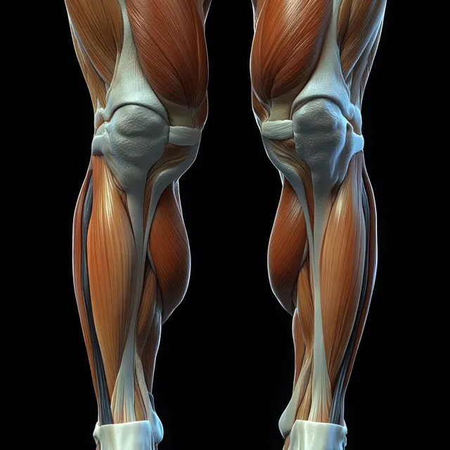
Anatomical illustration showing quadriceps and hamstrings muscles on both legs with highlighted knee joints.
Front anatomical view of human knees displaying femur, tibia, ligaments, tendons, and cartilage in vivid detail.
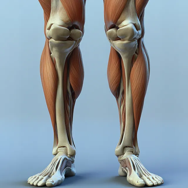
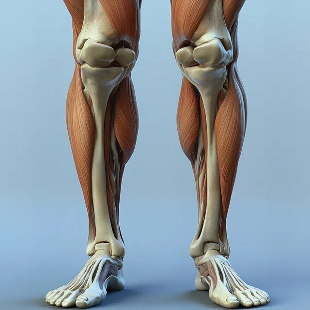
Illustrated legs show quadriceps muscles and detailed knee joints with exposed bone structure in a clinical setup.
3D anatomical illustration of a human skeleton with biceps brachii muscles highlighted on the upper arms.
3D skeleton torso with pectoralis minor highlighted orange and ribcage visible against a light teal background.
3D knee joint model with highlighted bones and cartilage resting on a smooth blue surface.