Arthritis images

Explore stock images tagged arthritis
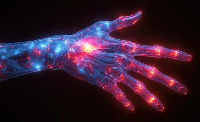
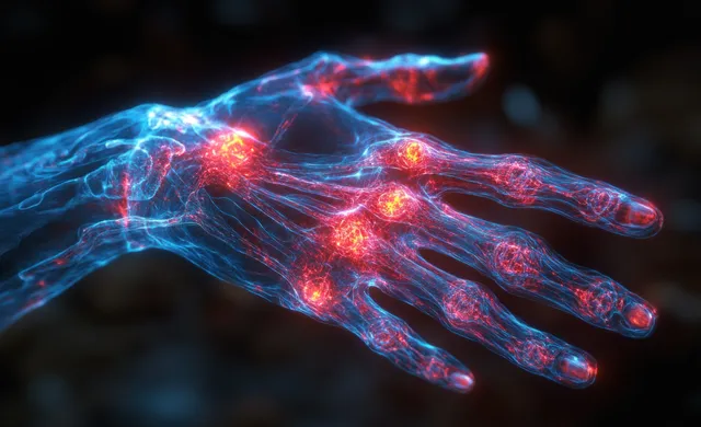
Neon 3D x-ray style hand and wrist with glowing carpal tunnel and joint pain highlights.
X-ray style illustration of a hand and wrist with glowing red and blue inflammation signals.
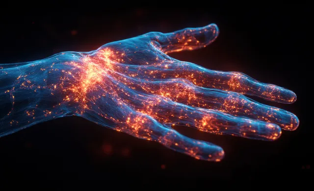
Digital wrist pain visualization with glowing x-ray hand over a dark background.
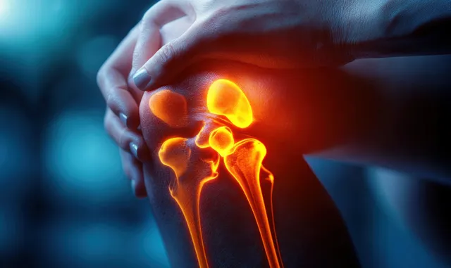
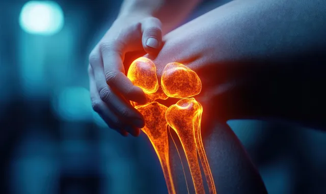
Hand holding an aching knee with a glowing knee joint and inflammation effect.
Neon blue and red X-ray style human figure highlights glowing joints and spine pain points.
A middle-aged Asian woman rests on a couch at home while holding her sore foot.
Hand gripping a painful knee with glowing bones overlay suggesting inflamed internal joint.
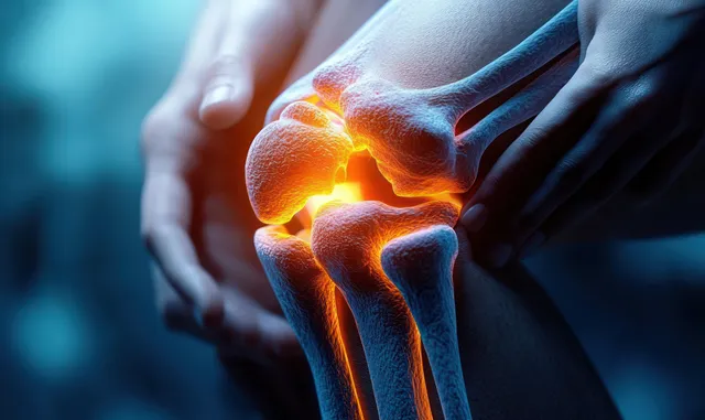
Hands hold a human knee as an orange glowing area marks inflammation or pain on the joint.
A hand presses a knee while glowing bone outlines indicate internal joint pain and injury.
A glowing neon anatomical illustration shows the shoulder and scapula musculature with highlighted bone structures.
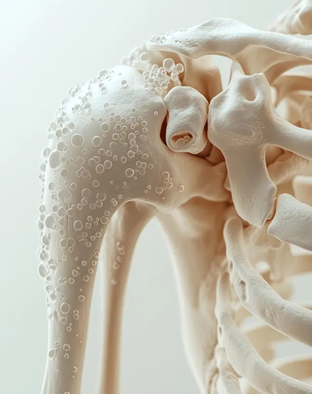
3D skeleton shoulder model with highlighted clavicle and scapula area suggesting painful inflammation.
3D render of anterior knee anatomy showing femur, patella, meniscus, cartilage, and surrounding ligaments on a dark background.
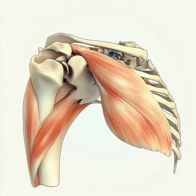
Side view anatomical illustration displaying shoulder muscles over the scapula and humerus with realistic soft shading.
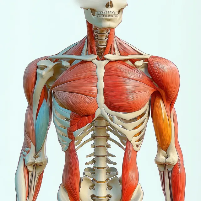
Front view 3D anatomy model showing human skeleton with red biceps brachii muscles highlighted.
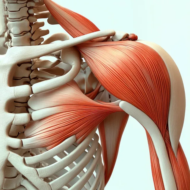
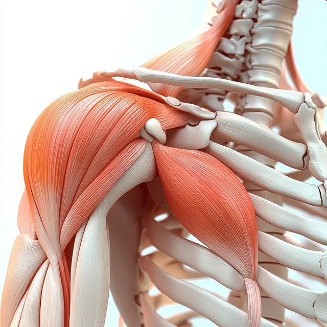
Anterior shoulder anatomy model showing highlighted deltoid muscle with visible clavicle, scapula, and rib cage.
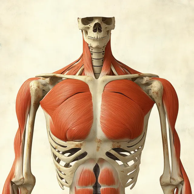
3D anatomical render of the pectoral muscles with tendon highlights and rib structure on a black background.
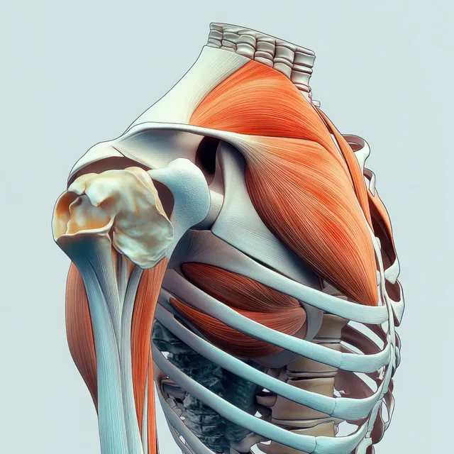
Side-view medical illustration of the shoulder joint with highlighted deltoid muscle over the ribcage.
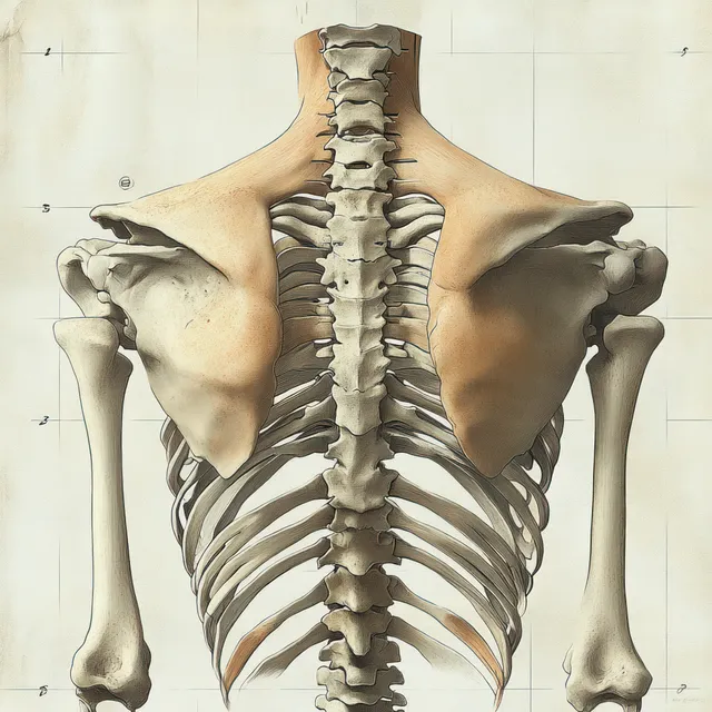
Back-view human skeleton illustration showing spine, ribcage, and shoulder blades against a light studio background.
3D anatomical view of the deltoid muscle on the shoulder with clavicle and scapula visible behind.
Red pectoralis major muscles are shown on an anterior human skeleton with labeled ribs, clavicle, and shoulder bones.
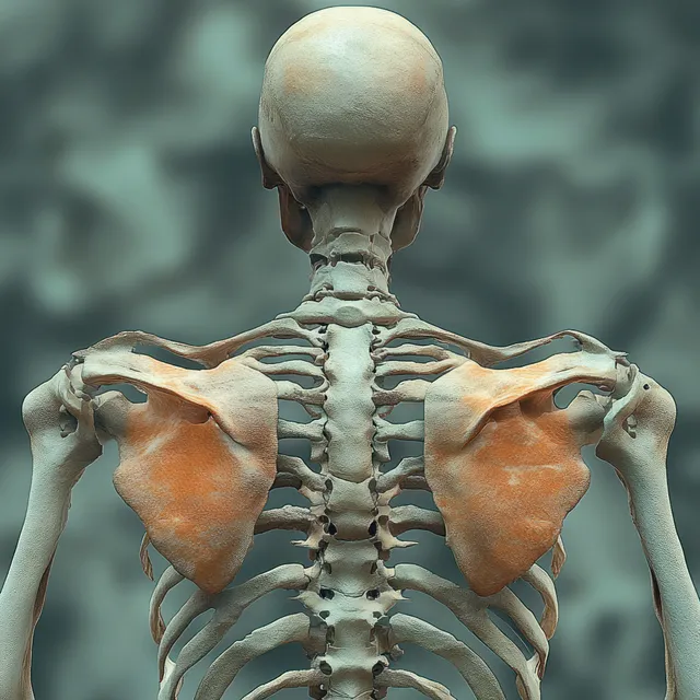
Rendered human skeleton viewed from behind with highlighted shoulder joints and supraspinatus muscles.
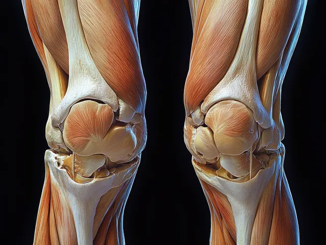
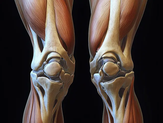
Symmetrical anatomical illustration of both knees showing femur and tibia with cartilage, meniscus, tendons, and ligaments on a dark background.
A man seated at a home office computer clutching his lower back in visible discomfort.
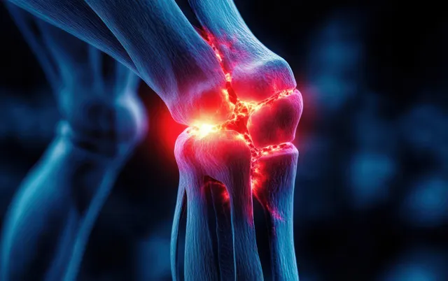
Digital knee joint model with glowing red inflamed areas around the joint and surrounding bones.