Cell Membrane images

Explore stock images tagged cell membrane
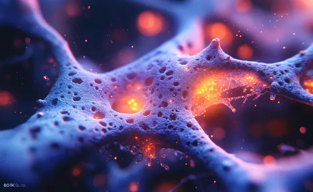
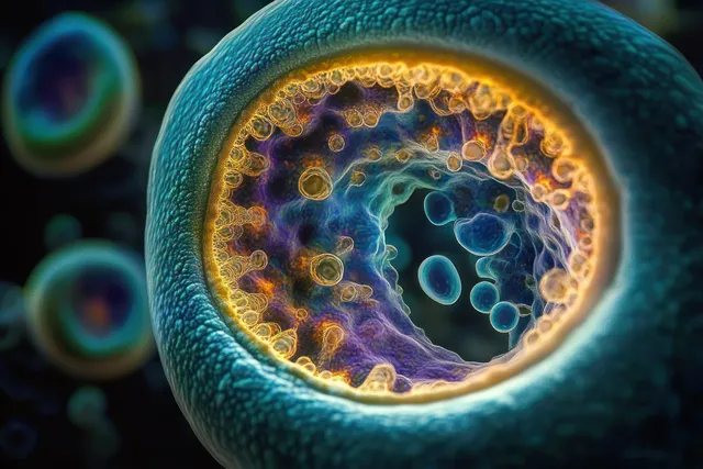
Curved cellular membrane with glowing yellow dots in a soft blue light close-up.
Glowing stem-cell membranes with orange neon organelle particles floating in a dark biotech lab background.
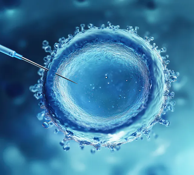
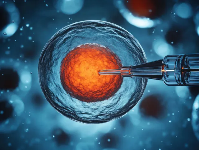
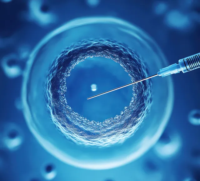
A blue single cell under a microscope is pierced by a fine injection needle, with surrounding particles in view.
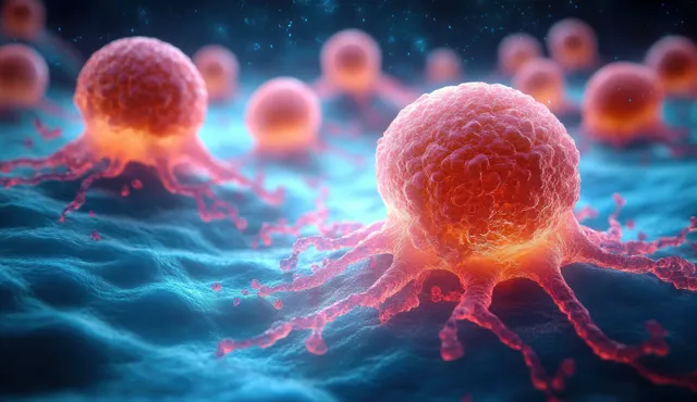
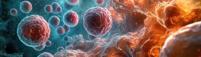
Glowing orange cancer cells with visible membrane tentacles float in a blue microscopic 3D background.
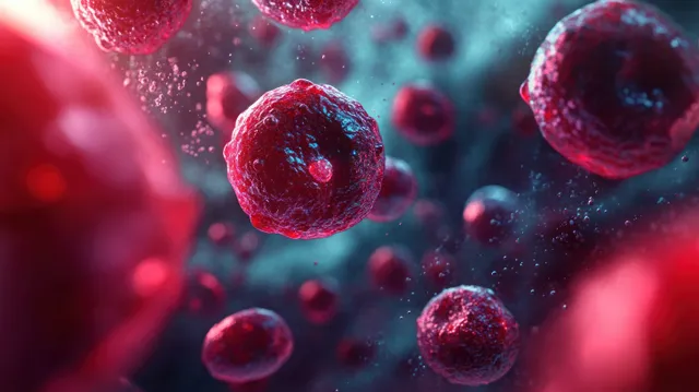
Microscopic red blood cells float in a red and blue fluid field with glowing highlights and detailed membranes.
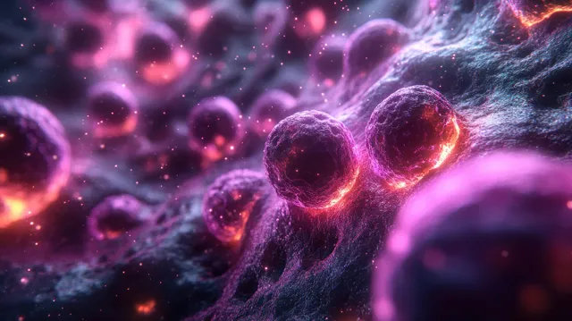
Glowing spherical cells float in a vibrant pink and purple background with visible internal structures.
Two glowing bacteria cells splitting in a dark microscopic environment, with blue membrane outlines and orange internal light.
A glowing purple spherical cell with several bright internal nucleoli floating on a clean background.
Red blood cells drift through a blue bloodstream background with glowing red highlights and fine cellular texture.
A microinjection needle pierces a cell nucleus inside a blue liquid medium with surrounding bubbles.
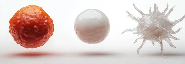
Three differently textured cells float on a white background: an orange sphere, a smooth white sphere, and a spiky cell.
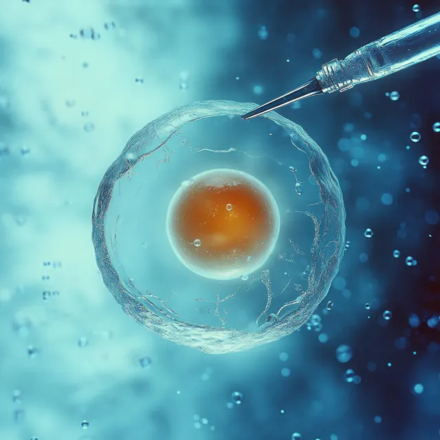
A glass pipette touches a transparent cell containing an orange nucleus.
Red blood cells move through a blood vessel in a microscopic view with swirling fluid around them.
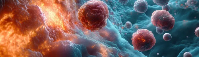
Textured spherical cells float in a fluid with contrasting blue and orange glowing light.
Red and orange textured spherical cells float among blue glowing microscopic vesicles in a dark, illuminated background.
Bioluminescent microbes glow inside a circular membrane in an undersea microscopic visualization.
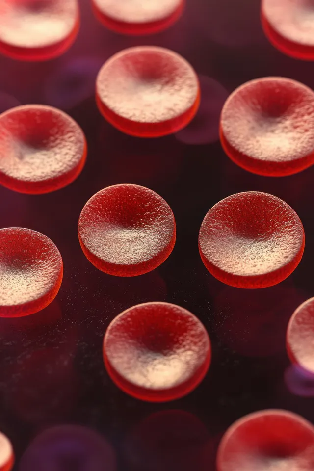
Glowing red disk-shaped cells with textured edges float against a dark background.
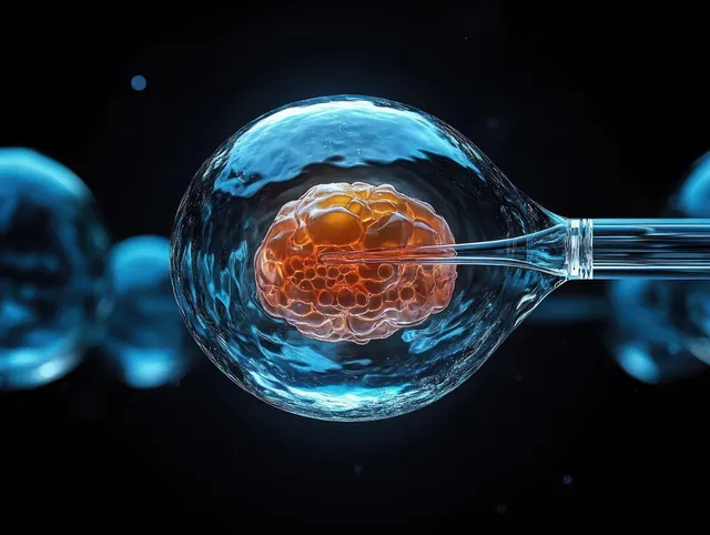
Microinjection needle pierces an orange cell while translucent blue membranes surround it in a microscopic view.
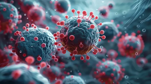
Stained microscopic view of a central virus surrounded by immune cells with red particles and blue cell membranes.
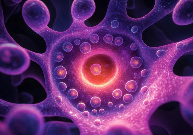
Glowing purple cell interior with vesicles around a bright central nucleus in a microscopic 3D view.
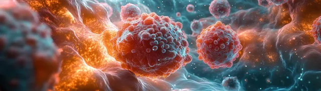
Close-up view of glowing cancer cells with textured surfaces interacting in a luminous fluid environment.
Microscopic embryo-like cells with glowing nuclei divide behind translucent membranes against a dark particle-filled background.
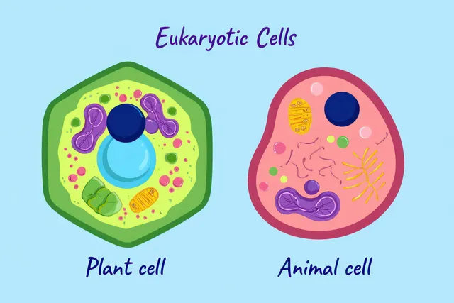
Two eukaryotic cell diagrams, plant and animal, display organelles like nucleus, chloroplasts, and mitochondria on a blue background.
A blue spherical cell is surrounded by blurred particles as a needle tip approaches for microinjection under a microscope.