Cross Sectional View images

Explore stock images tagged cross sectional view

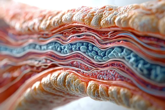
Close-up cross-section of layered biological tissue with cell clusters and wavy microtextures in warm red and blue tones.
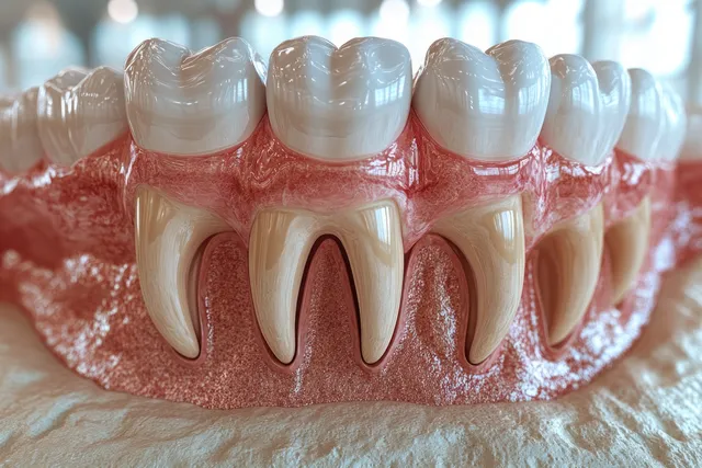
Dental cross-section model showing healthy enamel and inflamed gums with visible redness and swelling.
Medical anatomy illustration displays multiple heart angles beside a full-body figure with internal organs and blood vessel lines.
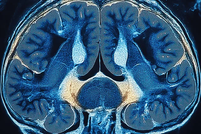
Horizontal cross-section image of the human brain with blue and beige internal structures and detailed folds.
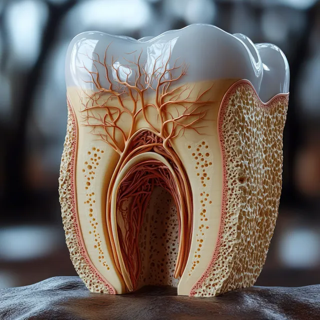
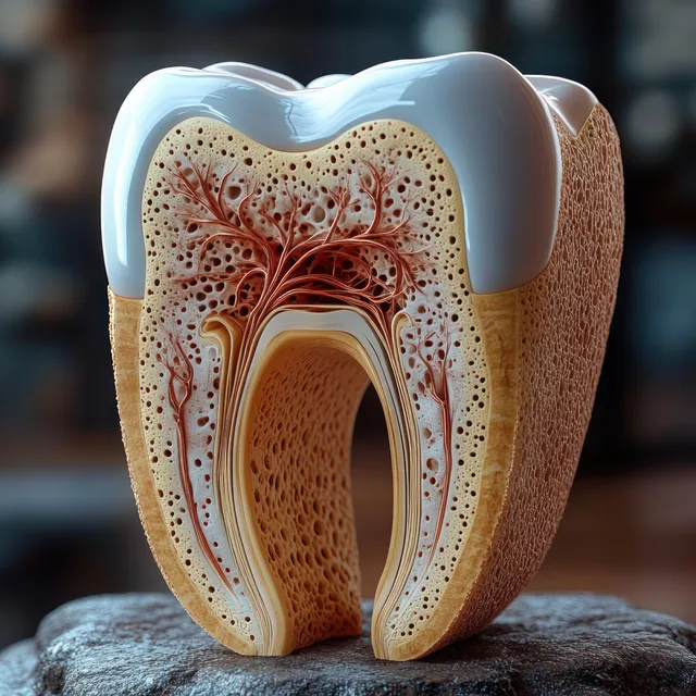
A human molar cross-section displays enamel, dentin, a pulp chamber with nerves and vessels, and porous root bone texture.
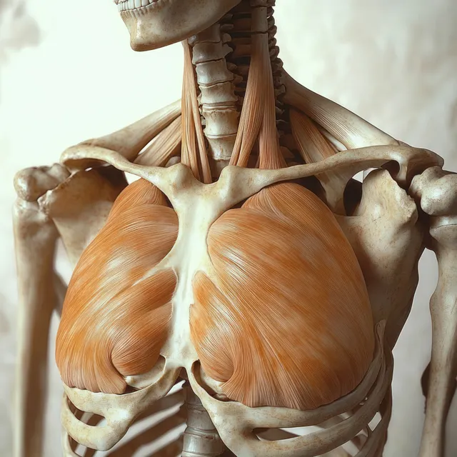
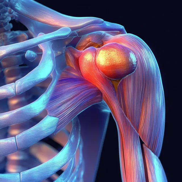
Renaissance anatomical view of a human skeleton with orange highlighted pectoralis major muscles across the chest.
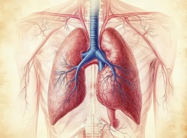
Medical illustration of human lungs, heart, and branching bronchial tree with blue blood vessels inside the ribcage.
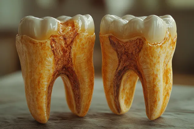
Two tooth cross-sections show one intact enamel surface and one decayed area exposing roots.
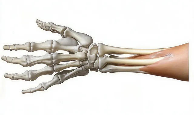
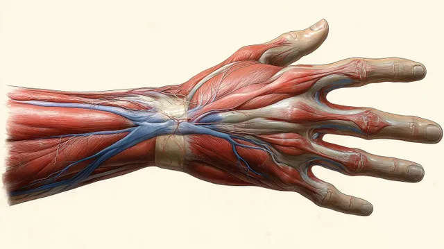
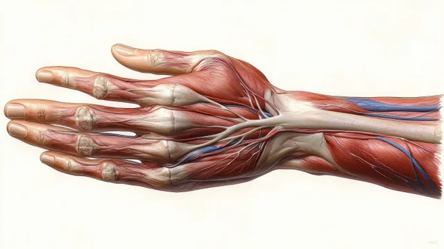
Layered anatomical illustration of a human hand showing bones, muscles, tendons, and skin from the wrist to fingers.
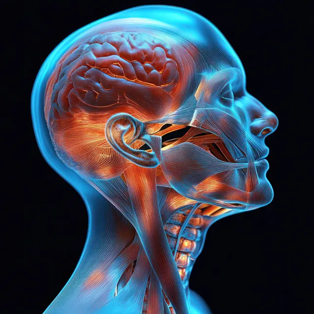
Profile view anatomical illustration of the human neck with highlighted muscles, nerves, and connective tissues.
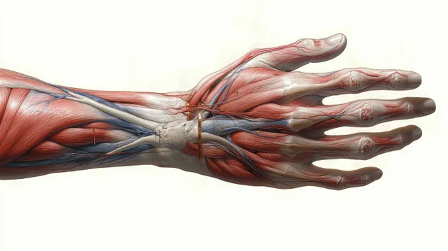
Ventral anatomical illustration of a female hand with visible muscles, arteries, veins, and tendons laid over bones.
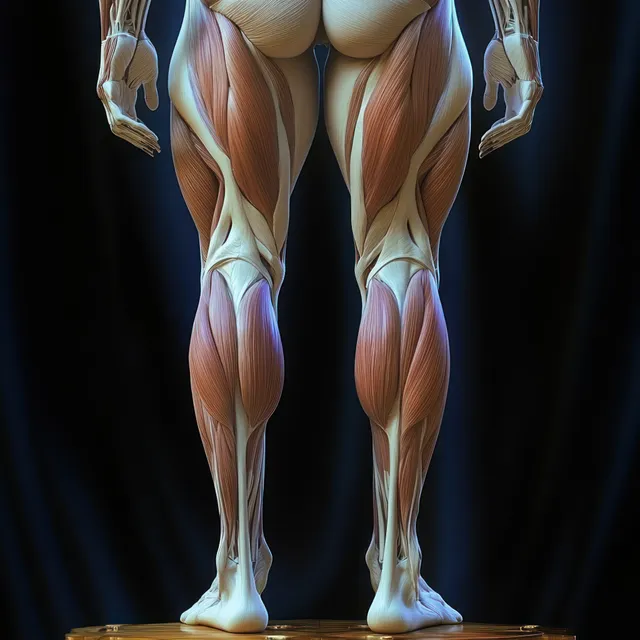
Female lower-body anatomical illustration displaying hip, thigh, and knee muscles with dark lighting and visible tendon lines.
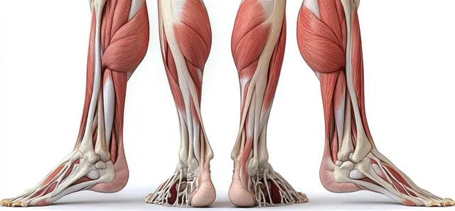
An anatomical cutaway illustration shows leg muscles and bones from front and back views on a white background.
Anatomical illustration of a female hand ventral view with visible muscles, tendons, arteries, and veins.
Ventral view hand anatomy illustration with visible muscle layers and branching blue blood vessels.
Detailed anatomical illustration of a human shoulder joint with deltoid and triceps highlighted against a white background.
Side profile anatomical diagram showing labeled human brain and spinal cord with visible internal regions.
Cross-sectional tooth model displays enamel, dentin, and pulp chamber with visible vessels and nerves.
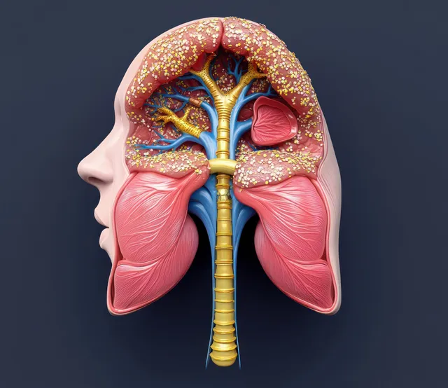
Cross-sectional anatomical illustration shows the human throat and nasal passages leading into the trachea and bronchi.
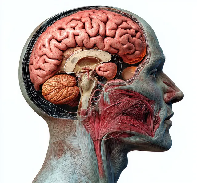
Side-view human head cutaway showing brain lobes, skull, and red nervous system tissues.