Detailed Muscle Fibers images

Explore stock images tagged detailed muscle fibers

3D anatomical render of the pectoral muscles with tendon highlights and rib structure on a black background.
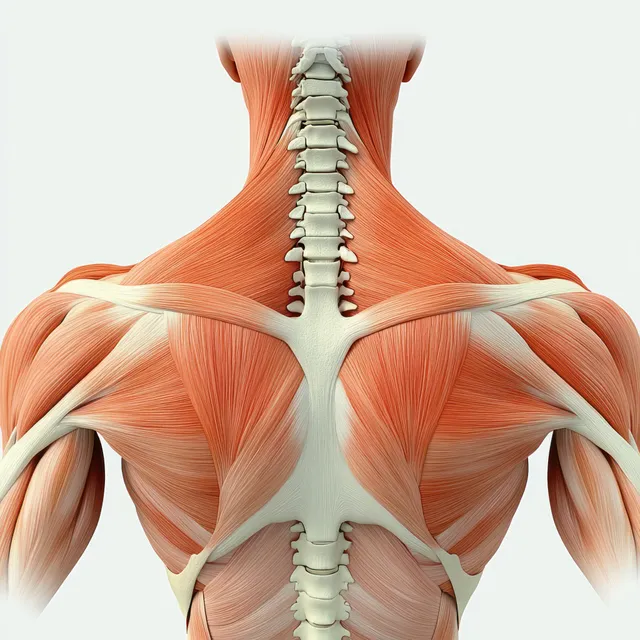
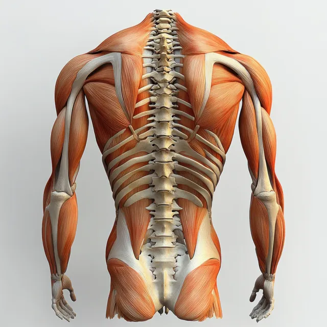
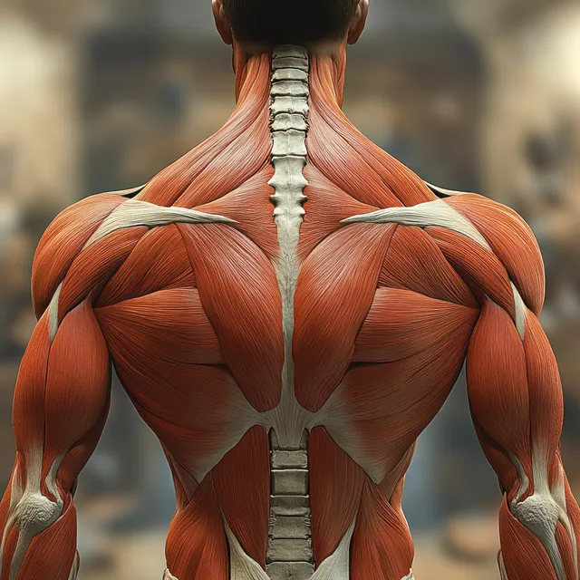
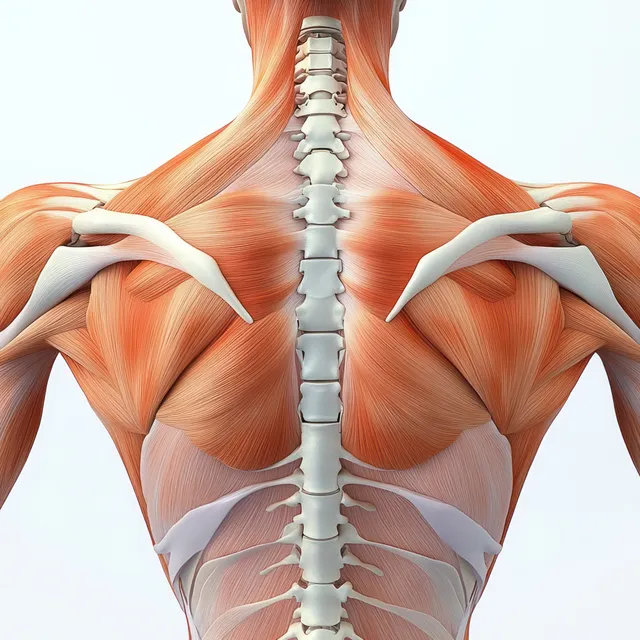
Posterior upper-back anatomy illustration with visible spine and shoulder muscles including rhomboid major and trapezius.
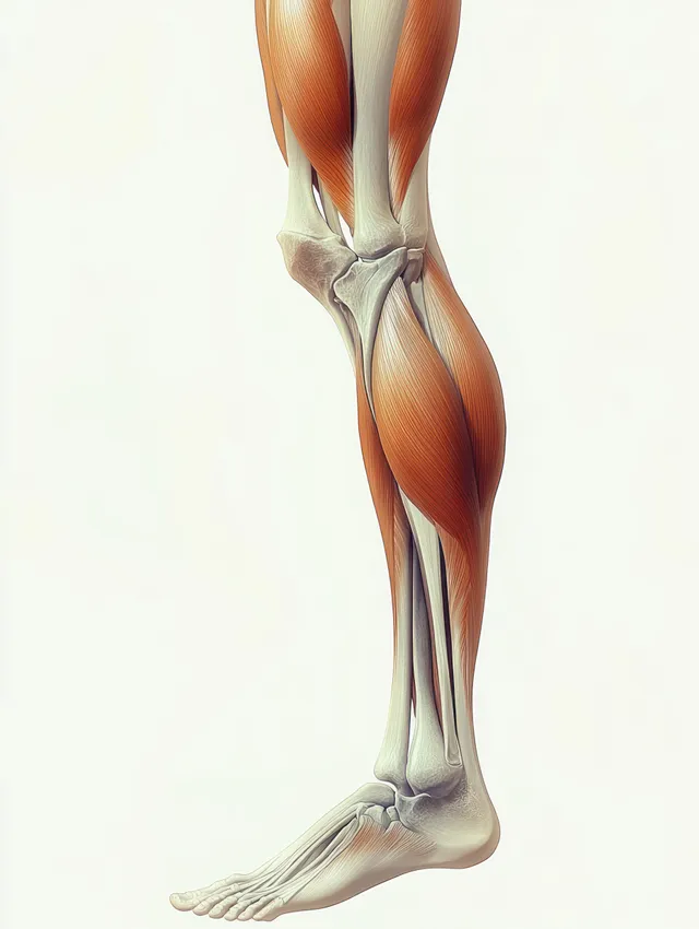
Side-view anatomical illustration of hamstring muscles on a human leg highlighting semitendinosus and biceps femoris longus.
Renaissance anatomy illustration highlights deltoid and triceps muscles with visible tendons and soft colored shading.
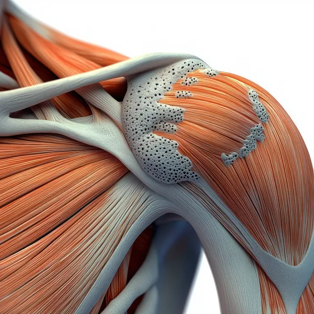
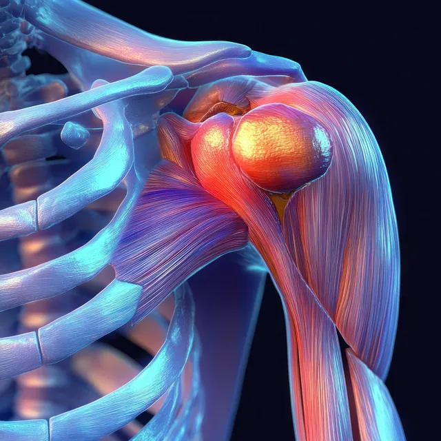
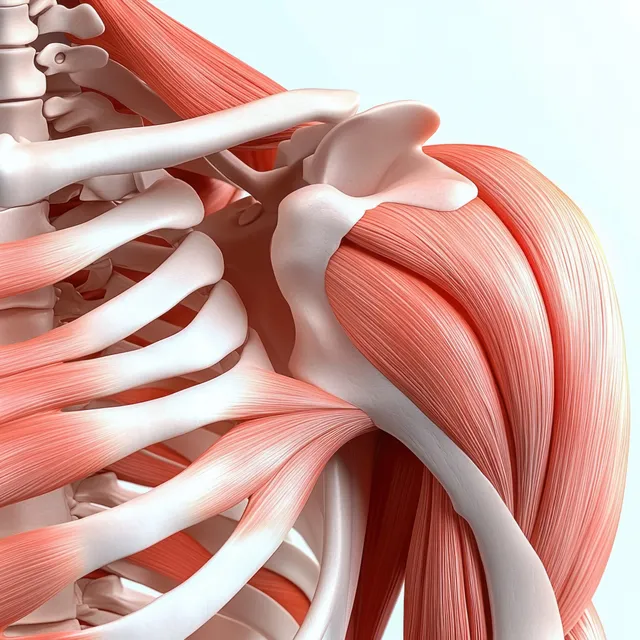
A 3D shoulder joint anatomical illustration shows humerus and scapula with visible tendon and muscle fiber detail.
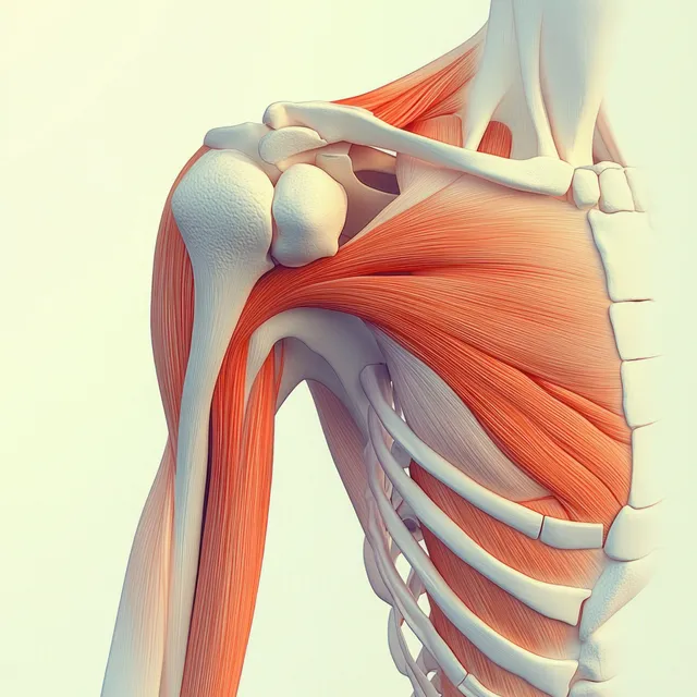
Detailed anatomical illustration of a human shoulder joint with deltoid and triceps highlighted against a white background.
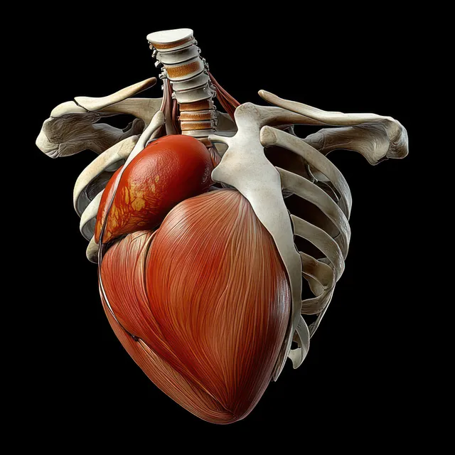
An anatomical render showing a red heart inside a visible ribcage with detailed muscle layers on a dark background.
Rear anatomical illustration highlighting erector spinae muscles along the spine with pelvis and lumbar vertebrae detail.
Anatomical shoulder diagram showing deltoid muscle bands and humerus bones with tendons and ligaments.
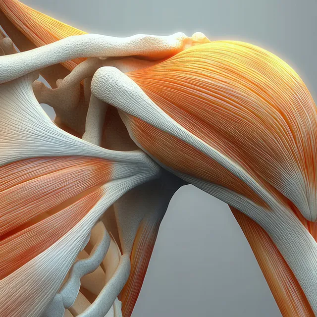
3D anatomical view of the human shoulder joint with orange triceps muscles wrapping the humerus.
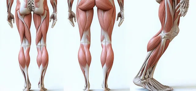
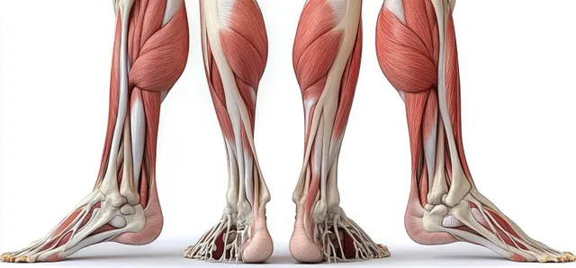
Front and back views of a male leg anatomy illustration with highlighted muscles, bones, tendons, and ligaments on a light background.
3D anatomical illustration of the human shoulder with exposed supraspinatus muscle over the humerus and scapula.
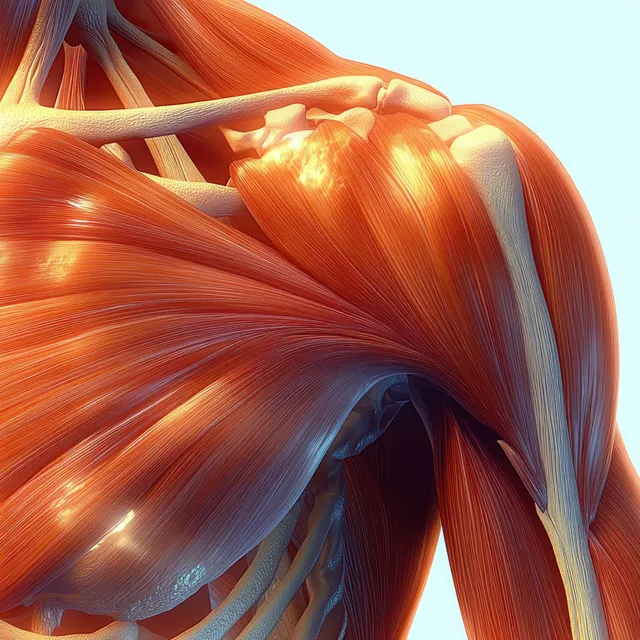
Anterior view anatomical illustration showing the deltoid muscle with clavicle and scapula bones and surrounding tissue layers.
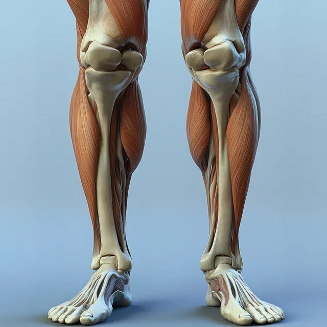
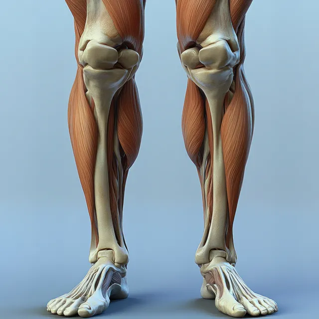
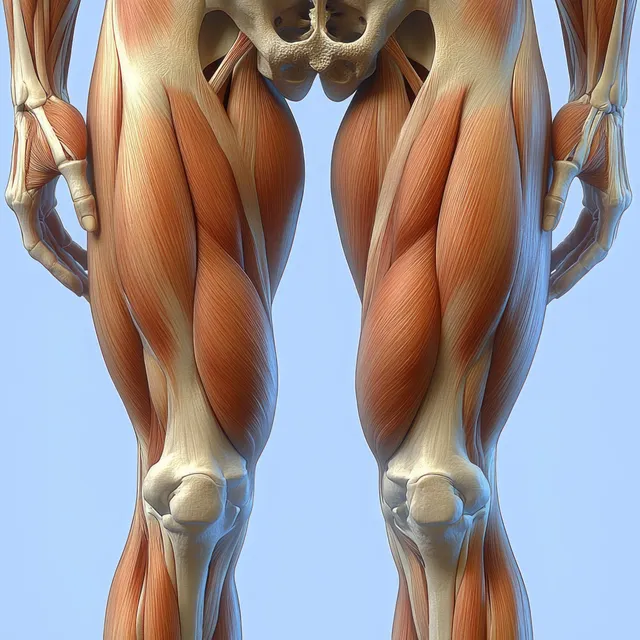
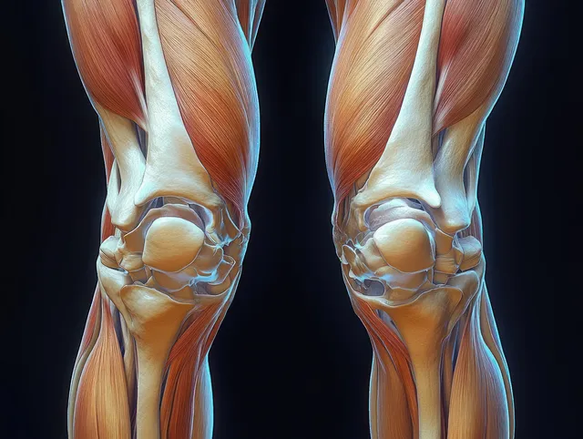
Illustrated legs show quadriceps muscles and detailed knee joints with exposed bone structure in a clinical setup.
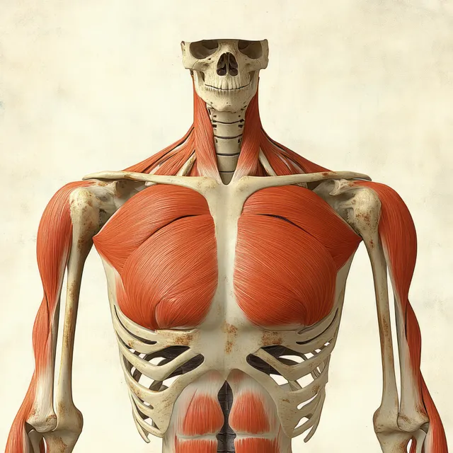
Red pectoralis major muscles are shown on an anterior human skeleton with labeled ribs, clavicle, and shoulder bones.
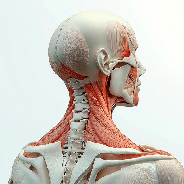
Anatomical rear-view figure with highlighted trapezius and neck muscles over a skeletal framework.
An anatomical cutaway illustration shows leg muscles and bones from front and back views on a white background.
Hyper-realistic view of male thighs and calves emphasizing quadriceps muscle groups and knee joints under blue backlight.
Rear view anatomical illustration reveals the trapezius muscles across the upper back and shoulders surrounding the spine.
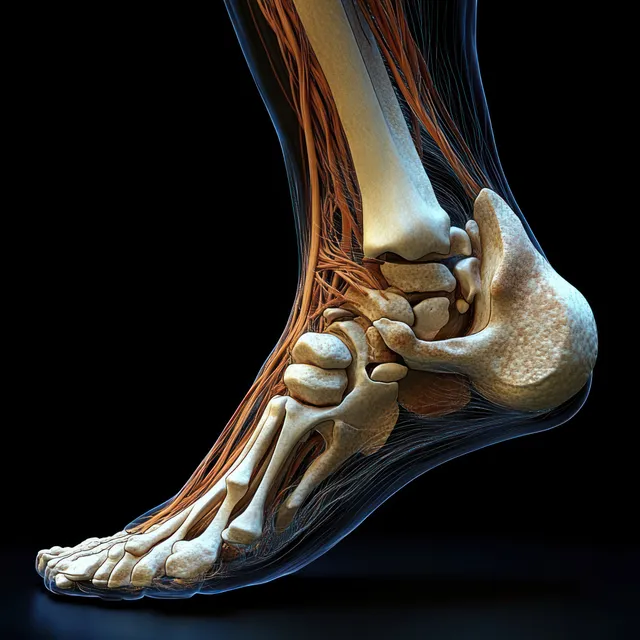
Medial x-ray style view of a human foot showing heel bones, ankle joints, and glowing tendons on a dark background.
Front anatomical view of human knees displaying femur, tibia, ligaments, tendons, and cartilage in vivid detail.
Illustrated upper back showing trapezius and rhomboid muscles with the spine centered.
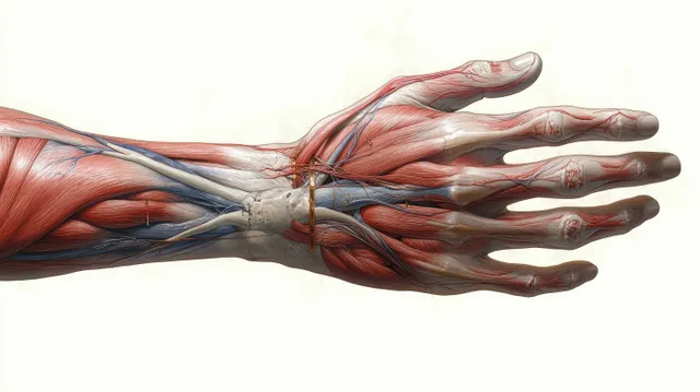
Ventral view hand anatomy illustration with visible muscle layers and branching blue blood vessels.