Educational Diagram images

Explore stock images tagged educational diagram

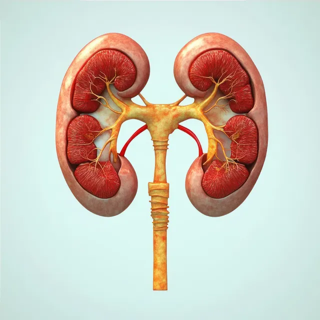
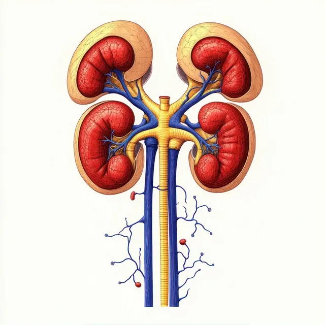
An anatomical kidney diagram displays renal arteries and veins with ureters on a light background.
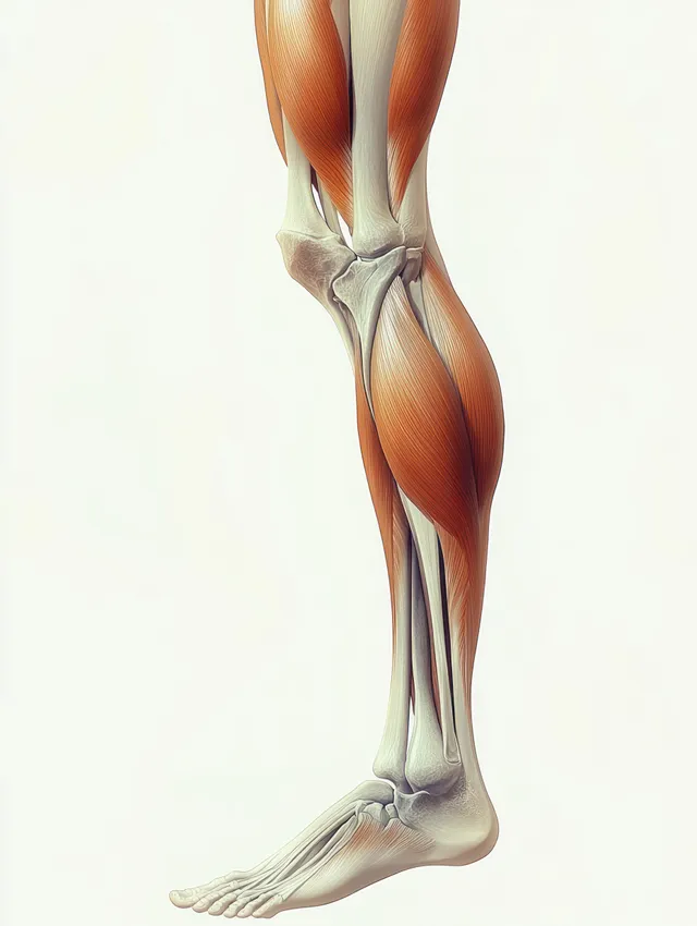
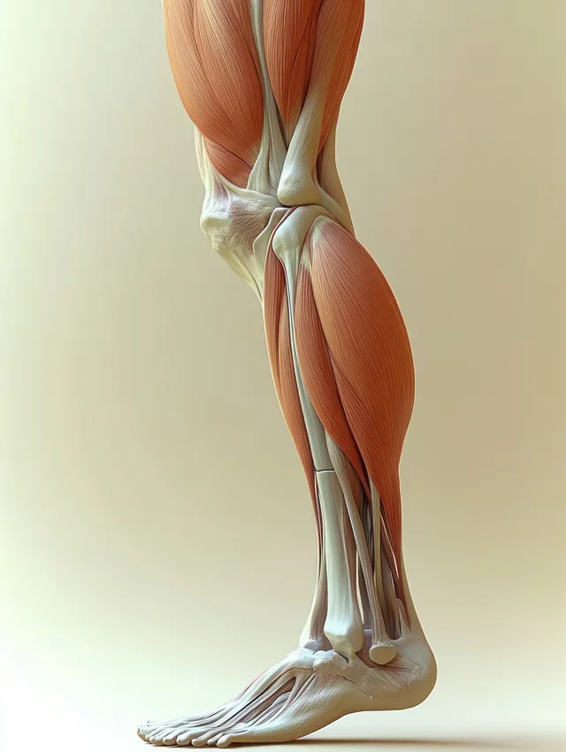
Side-view anatomical illustration of hamstring muscles on a human leg highlighting semitendinosus and biceps femoris longus.
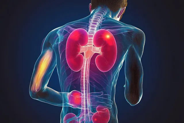
Transparent rear view of a human body with glowing kidneys, urinary tract, and visible spine and muscles against a dark background.
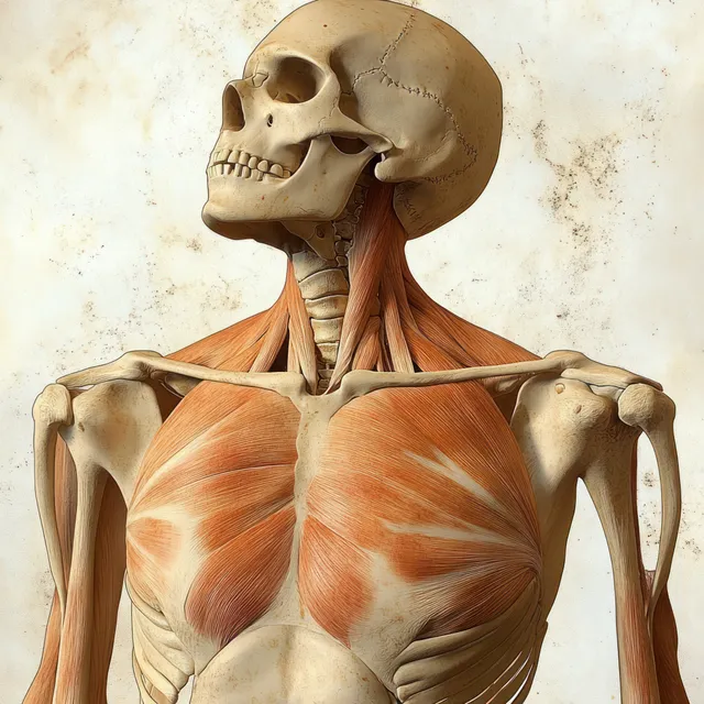
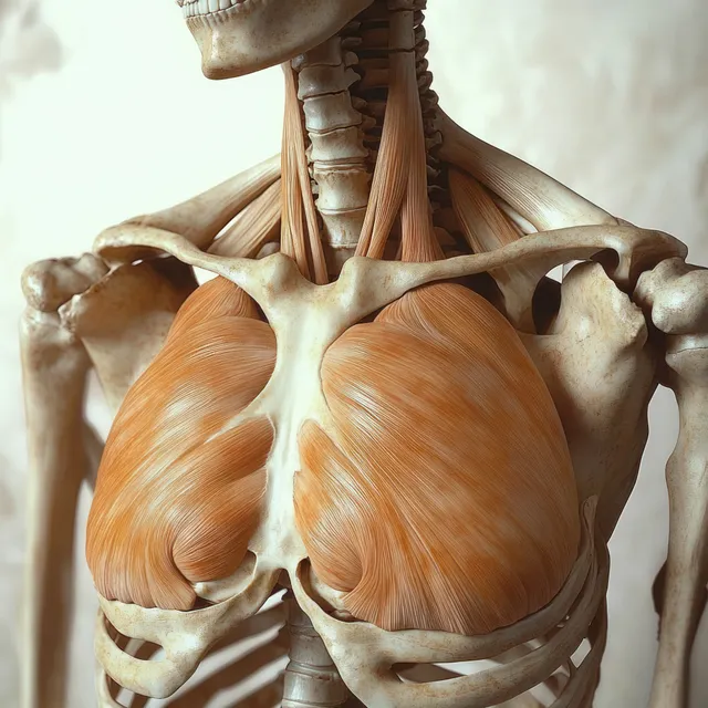
Skeleton model shown from the front with highlighted pectoralis major muscles and labeled chest anatomy.
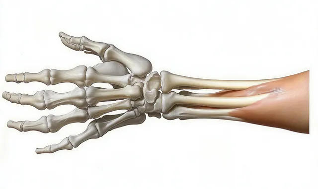
Layered anatomical illustration of a human hand showing bones, muscles, tendons, and skin from the wrist to fingers.
Four kidneys diagram showing ureters leading downward with connected blue blood vessels and internal renal regions.
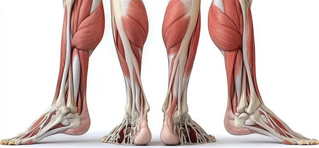
An anatomical cutaway illustration shows leg muscles and bones from front and back views on a white background.
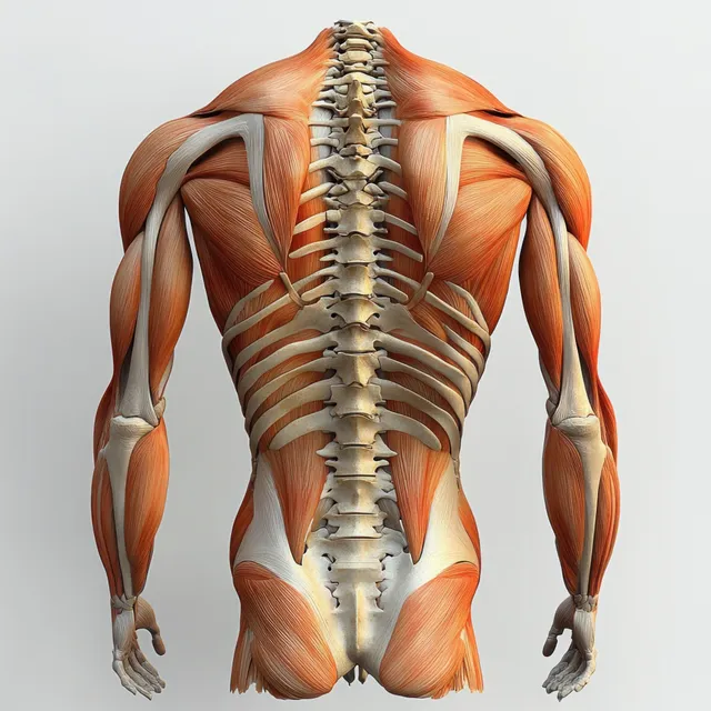
Rear anatomical illustration highlighting erector spinae muscles along the spine with pelvis and lumbar vertebrae detail.
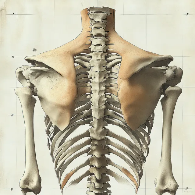
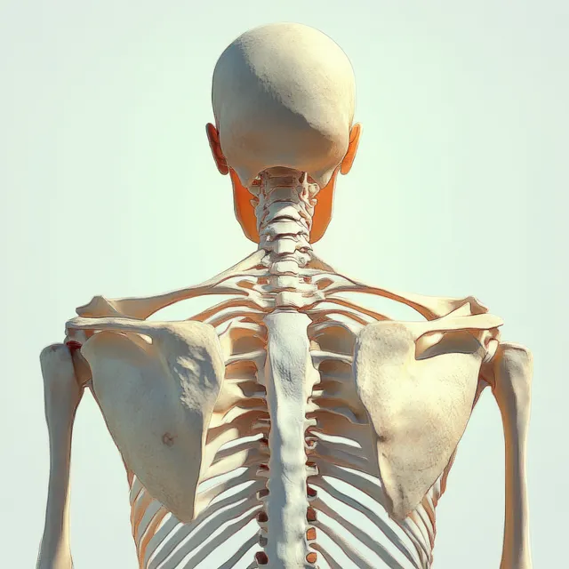
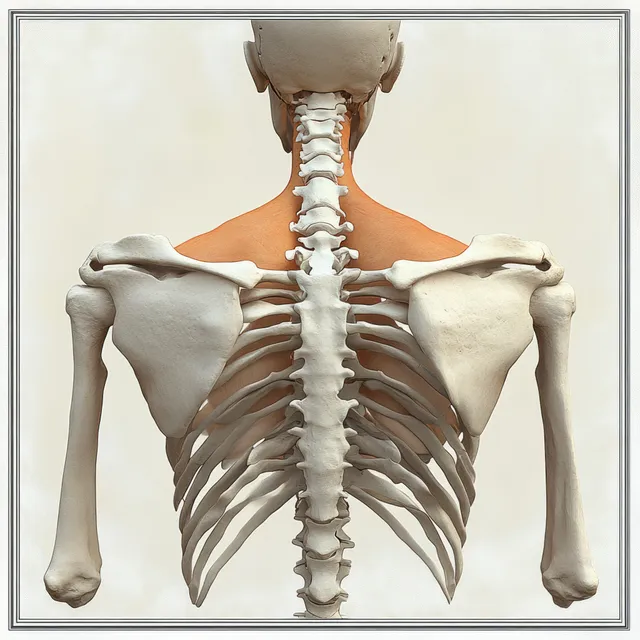
Back-view human skeleton illustration showing spine, ribcage, and shoulder blades against a light studio background.
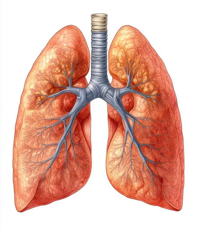
An anatomical illustration of human lungs with bronchi and visible vessels on a white background.
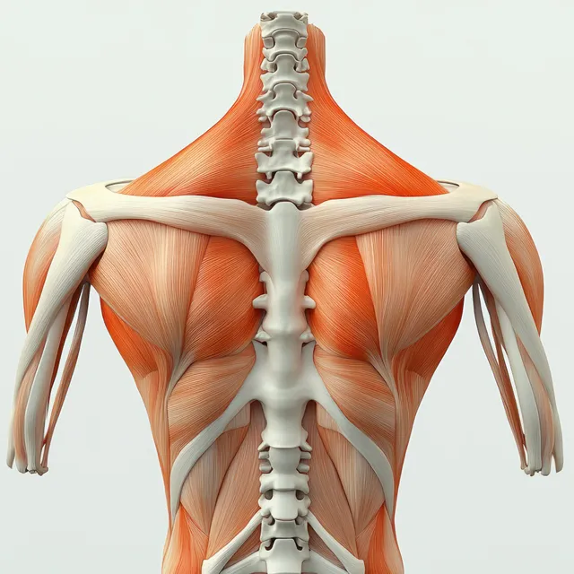
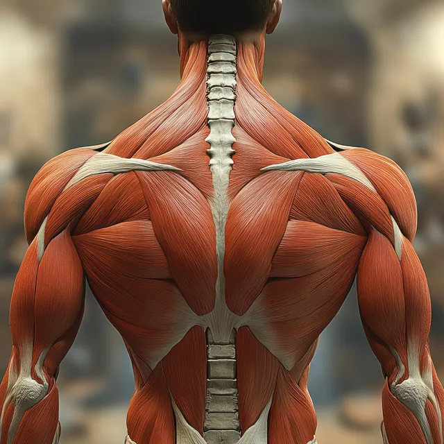
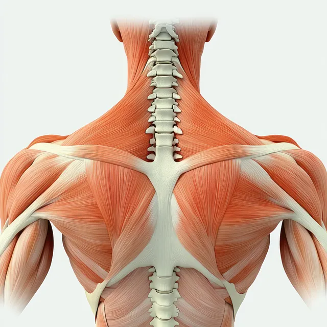
Rear view anatomical illustration displays trapezius and rhomboid muscles along the upper back over the spine.
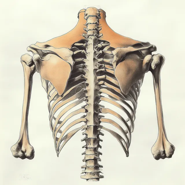
Rear view anatomical human skeleton showing spine and shoulder blades under soft studio light.
Renaissance anatomical view of a human skeleton with orange highlighted pectoralis major muscles across the chest.
Rear view anatomical illustration reveals the trapezius muscles across the upper back and shoulders surrounding the spine.
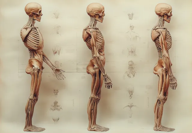
Anatomical illustration shows three views of a human figure’s skeleton and muscles standing against an off-white background.
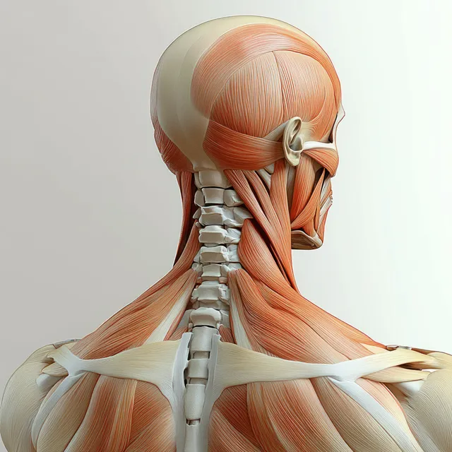
Rear view anatomical 3D illustration of the neck and trapezius muscles over the vertebrae.
Empty classroom interior with a wooden floor and a large chalkboard showing faint diagrams and chalk writing.
Anatomical illustration of a human skeleton torso highlighting the spine and shoulder blades under soft lighting.
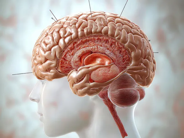
Anatomical profile illustration of a human brain with highlighted pineal gland, thalamus, and hypothalamus.
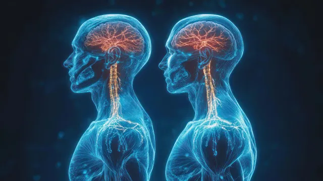
Two human side-profile figures display glowing brain and spinal cord neural pathways on a dark background.
Colorful DNA helix with gradient segments and thin connecting lines on a light background.
Rear view anatomical illustration of a human skeleton showing spine and shoulder blades with rhomboid minor emphasis.
Lateral view anatomical illustration of the hamstring muscles and knee joint with highlighted thigh tendons.
Posterior upper-back anatomy illustration with visible spine and shoulder muscles including rhomboid major and trapezius.