Labeled Diagram images

Explore stock images tagged labeled diagram

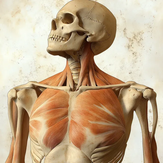
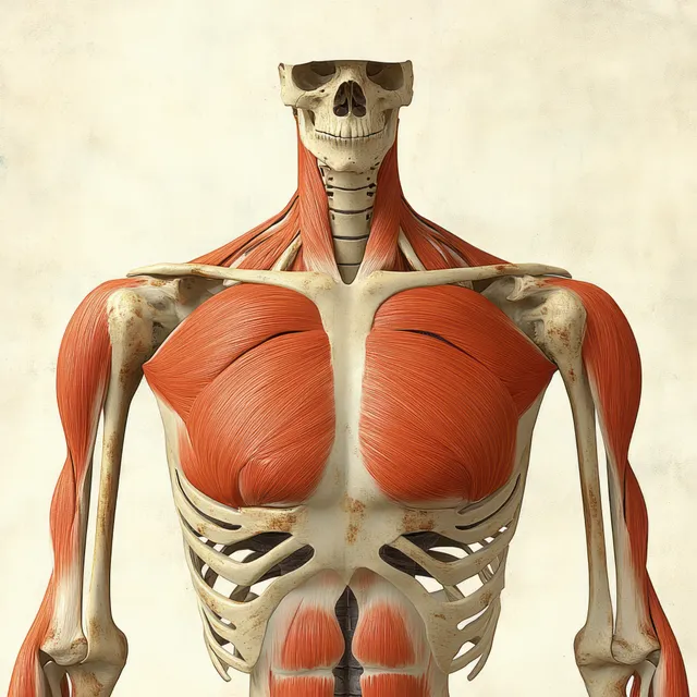
Skeleton model shown from the front with highlighted pectoralis major muscles and labeled chest anatomy.
Red pectoralis major muscles are shown on an anterior human skeleton with labeled ribs, clavicle, and shoulder bones.
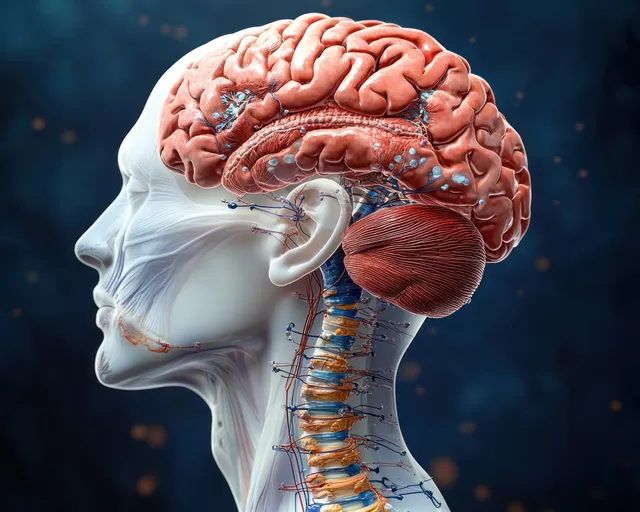
Side profile anatomical diagram showing labeled human brain and spinal cord with visible internal regions.
Labeled shoulder joint anatomy illustration showing clavicle, scapula, humerus, and ligaments on a white background.
A labeled profile view shows the human brain and spinal cord with translucent internal structures and vertebrae.
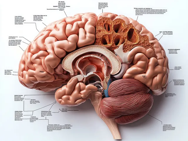
Labeled sagittal brain diagram showing frontal lobe and cerebellum in detailed anatomical section.
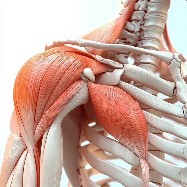
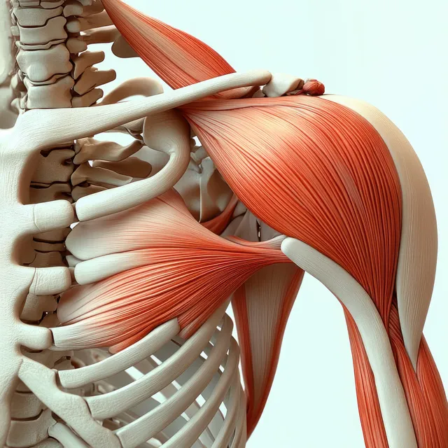
3D anatomical view of the deltoid muscle on the shoulder with clavicle and scapula visible behind.
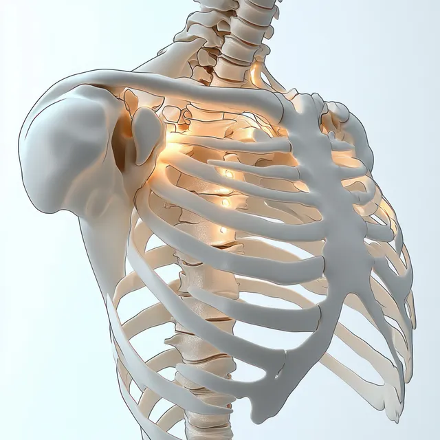
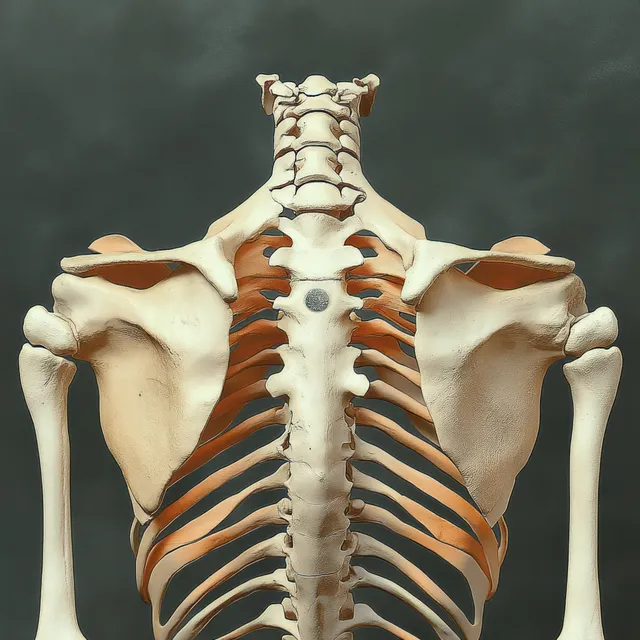
Rear view human skeleton model focusing on the spine, shoulder blades, and ribcage.
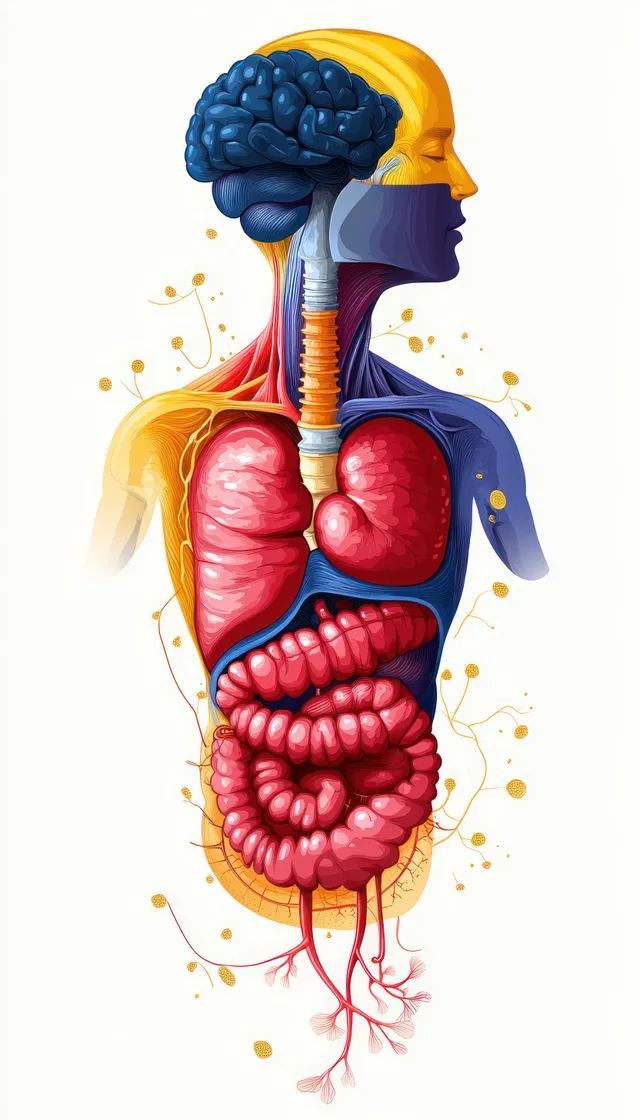
Color-coded cross-section illustration of the human digestive system showing stomach, intestines, colon, liver, and pancreas.
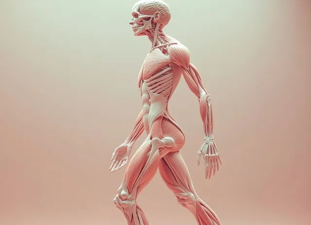
3D human figure anatomy view with transparent muscles and skeleton, showing labeled blood vessels on a soft background.
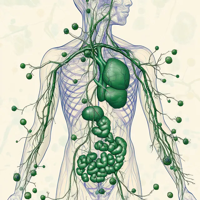
Green lymphatic vessels and clustered lymph nodes overlay the chest and abdomen on a female body diagram.
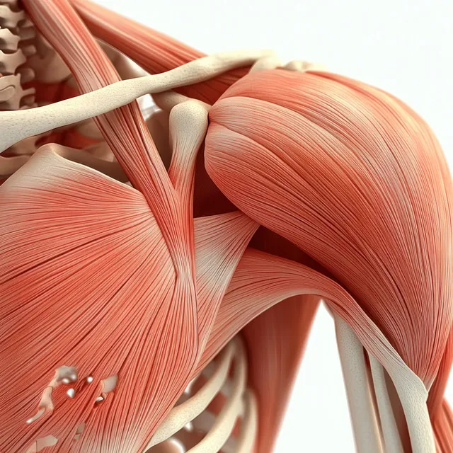
Anterior shoulder anatomy model showing highlighted deltoid muscle with visible clavicle, scapula, and rib cage.
Three pastel pyramid icons labeled Level A and Level B with soft shading on a white background.
Two eukaryotic cell diagrams, plant and animal, display organelles like nucleus, chloroplasts, and mitochondria on a blue background.
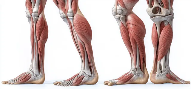
Front and back cutaway views of human legs with visible thigh and calf muscles and leg bones on a white background.
Renaissance anatomy illustration highlights deltoid and triceps muscles with visible tendons and soft colored shading.
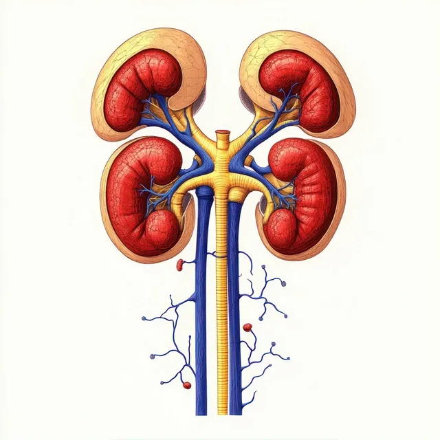
Four kidneys diagram showing ureters leading downward with connected blue blood vessels and internal renal regions.
Anterior shoulder anatomy illustration highlighting the deltoid muscle, tendons, and surrounding bones.
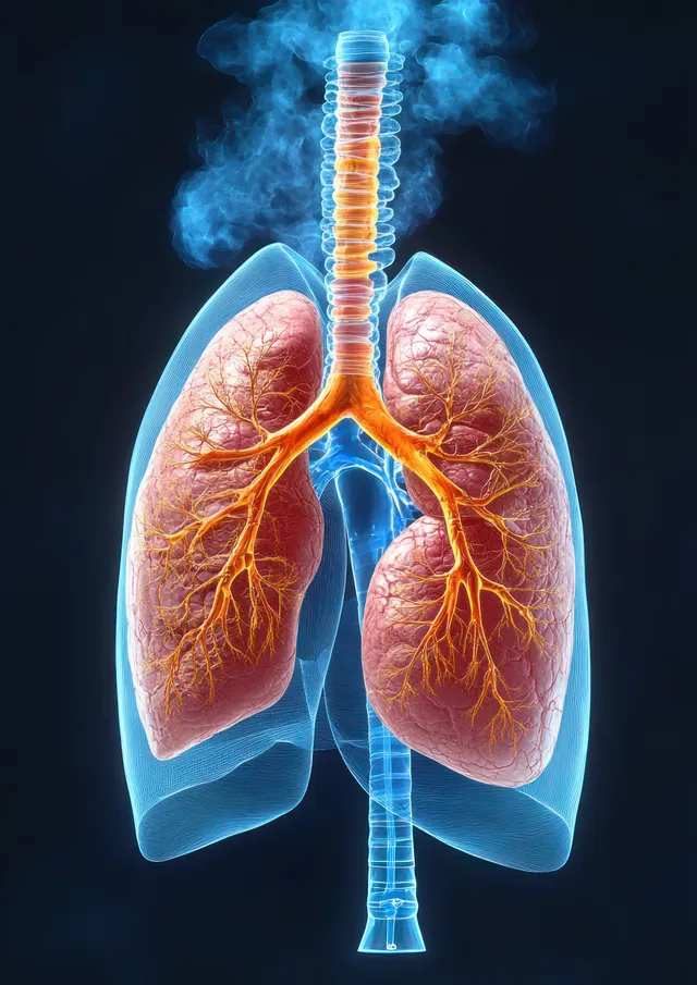
Glowing cross-section illustration of the human respiratory system with visible trachea, bronchi, and lungs inside the chest.
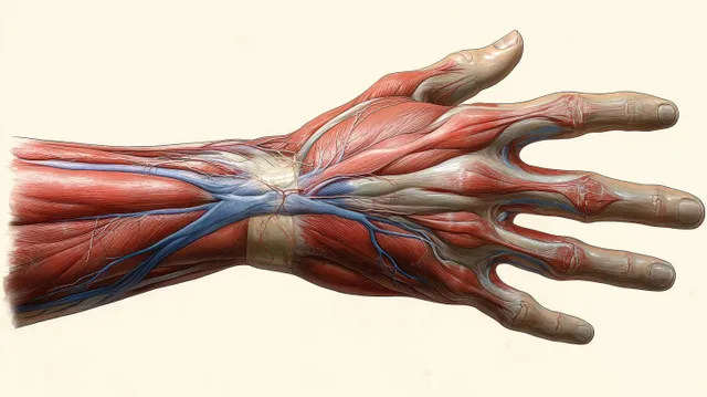
Ventral anatomical illustration of a female hand with visible muscles, arteries, veins, and tendons laid over bones.
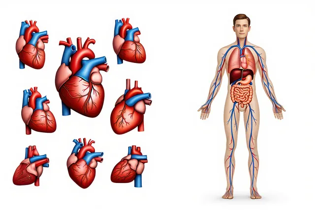
Medical anatomy illustration displays multiple heart angles beside a full-body figure with internal organs and blood vessel lines.
Glowing 3D digestive system anatomy with light particles showing esophagus, stomach, intestines, liver, and pancreas.
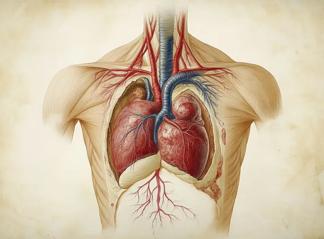
Renaissance anatomical cutaway of a human heart and circulatory vessels with a branching bronchial tree above.
Blue CAD-style ventilation blueprint displays metallic pipes with fittings and connected duct lines.