Musculoskeletal images

Explore stock images tagged musculoskeletal
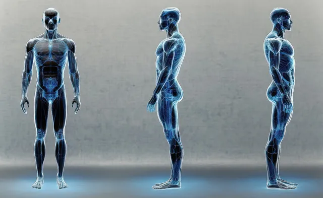
Three transparent male figures show front and side musculoskeletal anatomy on a textured gray background.
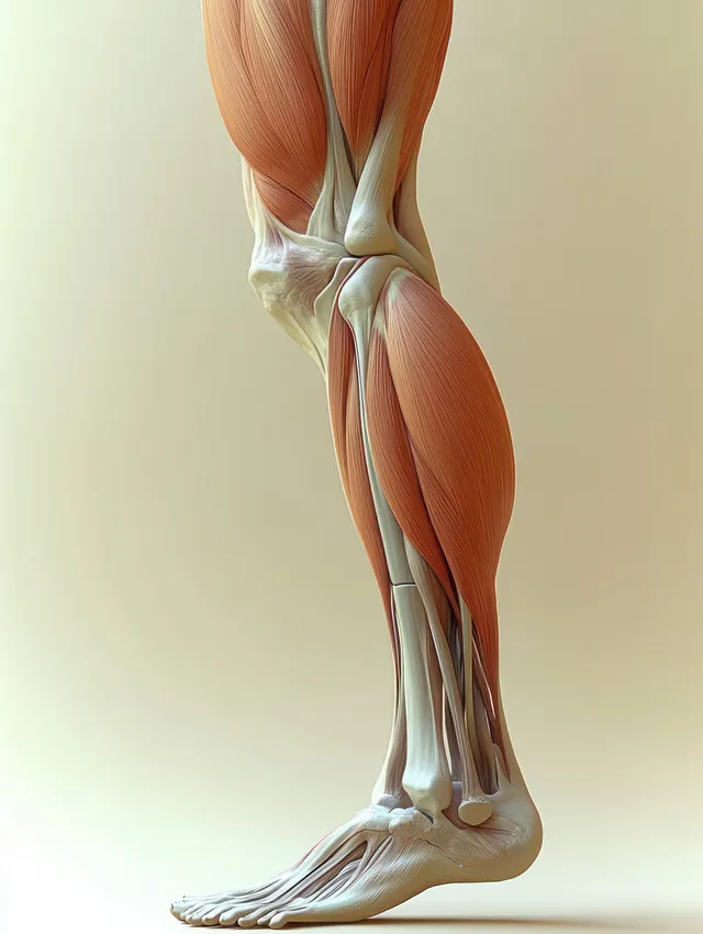
Lateral view anatomical illustration of the hamstring muscles and knee joint with highlighted thigh tendons.
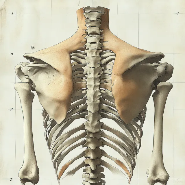
Back-view human skeleton illustration showing spine, ribcage, and shoulder blades against a light studio background.
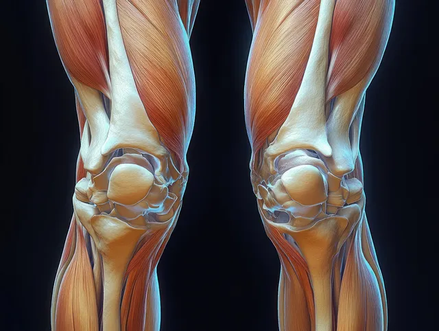
Front anatomical view of human knees displaying femur, tibia, ligaments, tendons, and cartilage in vivid detail.
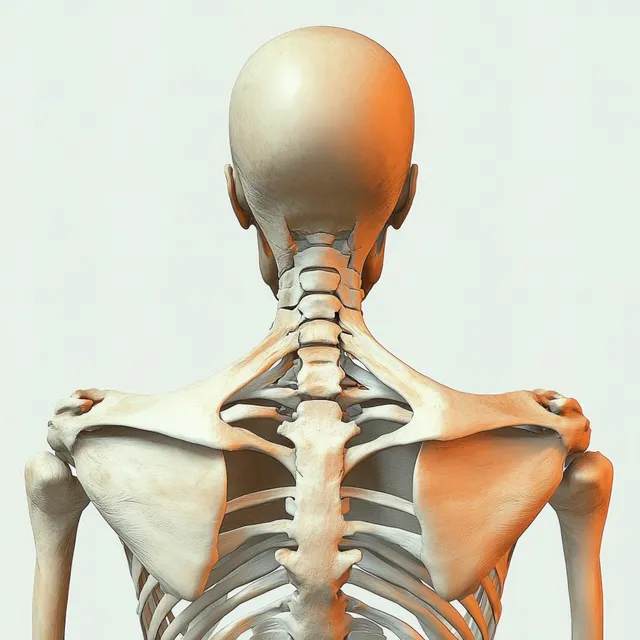
Rear view 3D anatomical illustration of a human skeleton showing spine, scapulae, and shoulders.
3D anatomical illustration of a human skeleton with biceps brachii muscles highlighted on the upper arms.
Translucent digital human anatomy shows glowing lungs and trachea with DNA helix strands behind it.
Translucent upper body medical illustration with a spine and red glowing vertebrae indicating inflammation.
Hands hold a human knee as an orange glowing area marks inflammation or pain on the joint.
A hand presses a knee while glowing bone outlines indicate internal joint pain and injury.
A hand holds a knee as glowing internal bones show beneath the skin on a blue background.
Hand holding an aching knee with a glowing knee joint and inflammation effect.
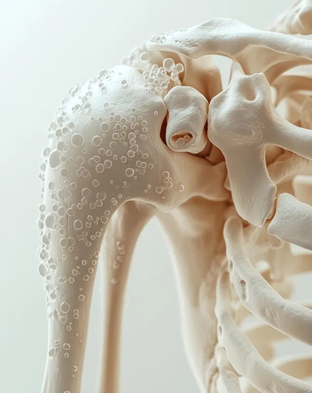
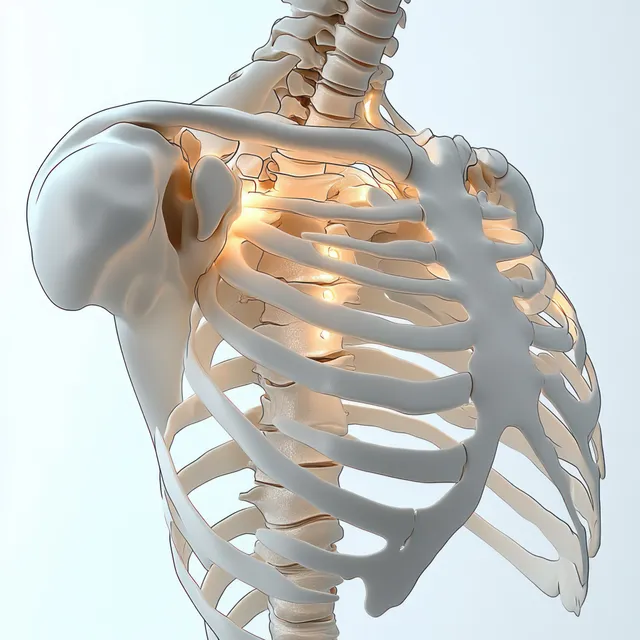
3D skeleton shoulder model with highlighted clavicle and scapula area suggesting painful inflammation.
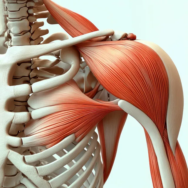
Anterior shoulder anatomy model showing highlighted deltoid muscle with visible clavicle, scapula, and rib cage.
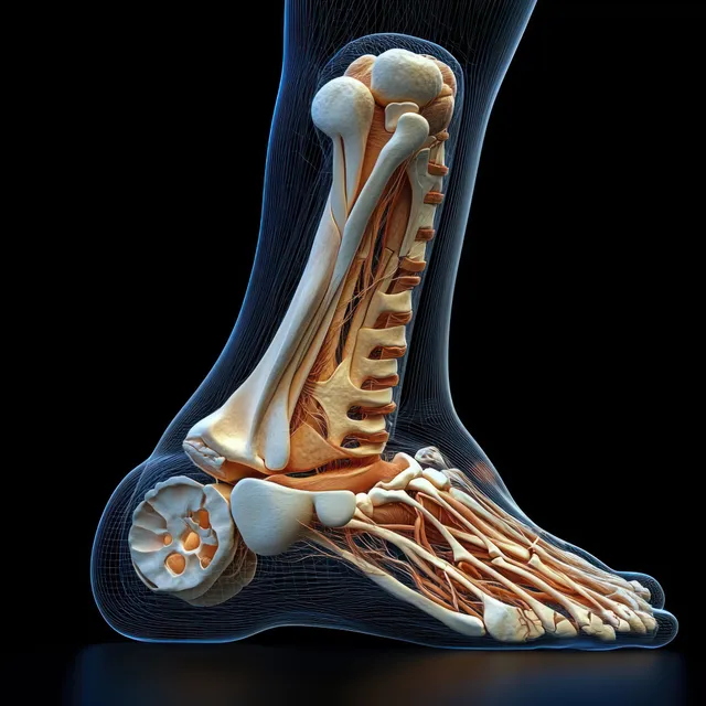
Surreal anatomical illustration of a human foot with exposed bones and tendons under dramatic rim lighting.
3D anatomical render of the pectoral muscles with tendon highlights and rib structure on a black background.
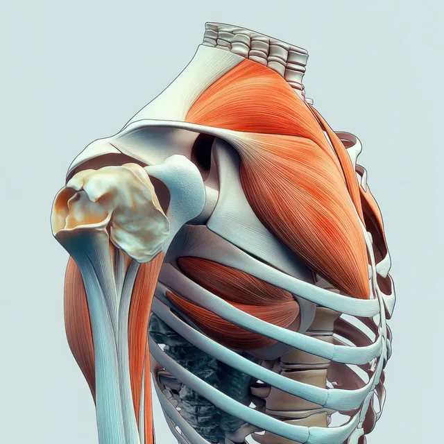
Side-view medical illustration of the shoulder joint with highlighted deltoid muscle over the ribcage.
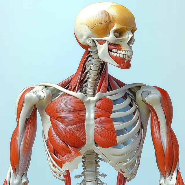
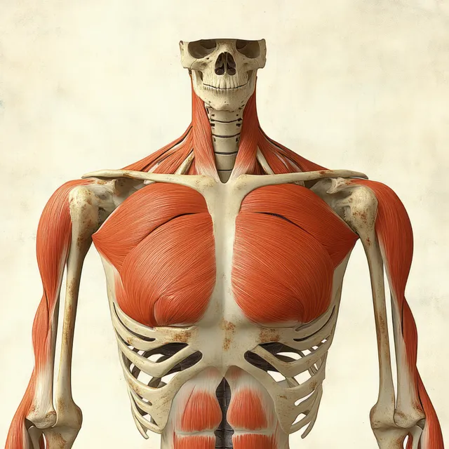
Red pectoralis major muscles are shown on an anterior human skeleton with labeled ribs, clavicle, and shoulder bones.
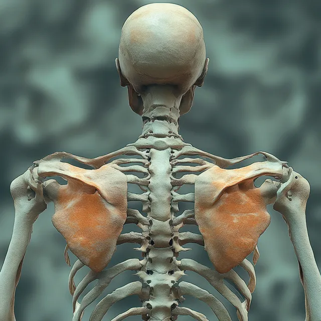
Rendered human skeleton viewed from behind with highlighted shoulder joints and supraspinatus muscles.
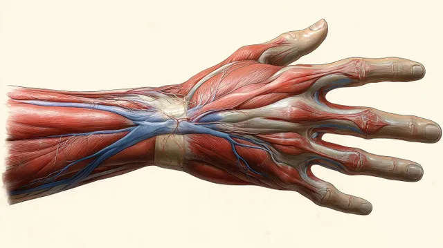
Ventral anatomical illustration of a female hand with visible muscles, arteries, veins, and tendons laid over bones.
Labeled shoulder joint anatomy illustration showing clavicle, scapula, humerus, and ligaments on a white background.
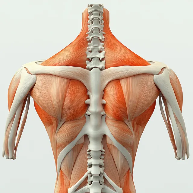
Rear view anatomical illustration displays trapezius and rhomboid muscles along the upper back over the spine.
Symmetrical anatomical illustration of both knees showing femur and tibia with cartilage, meniscus, tendons, and ligaments on a dark background.
Runner holding her aching knee on an asphalt road with a glowing pain spot near the joint.
Musculoskeletal images | StockPico