Posterior Anatomical View images

Explore stock images tagged posterior anatomical view
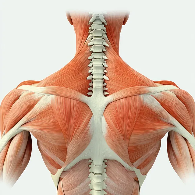
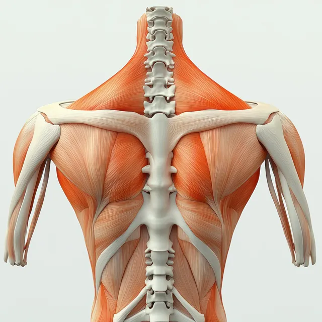
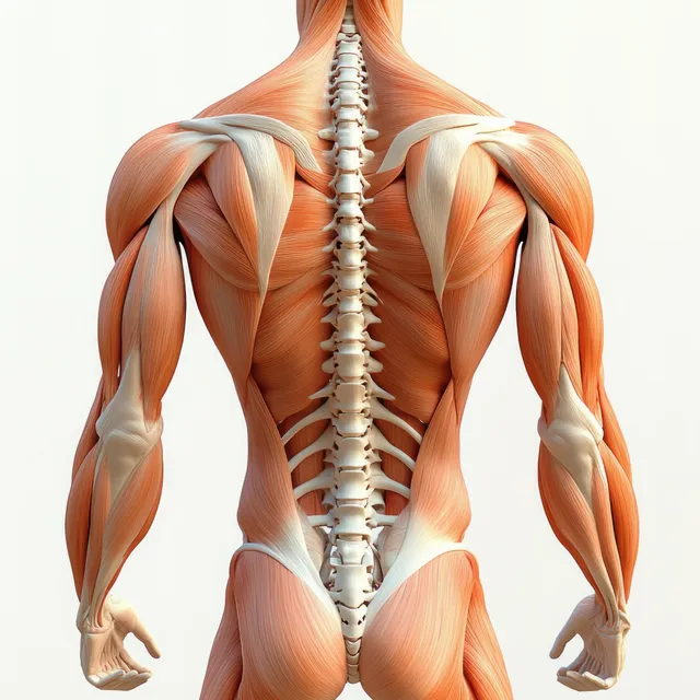
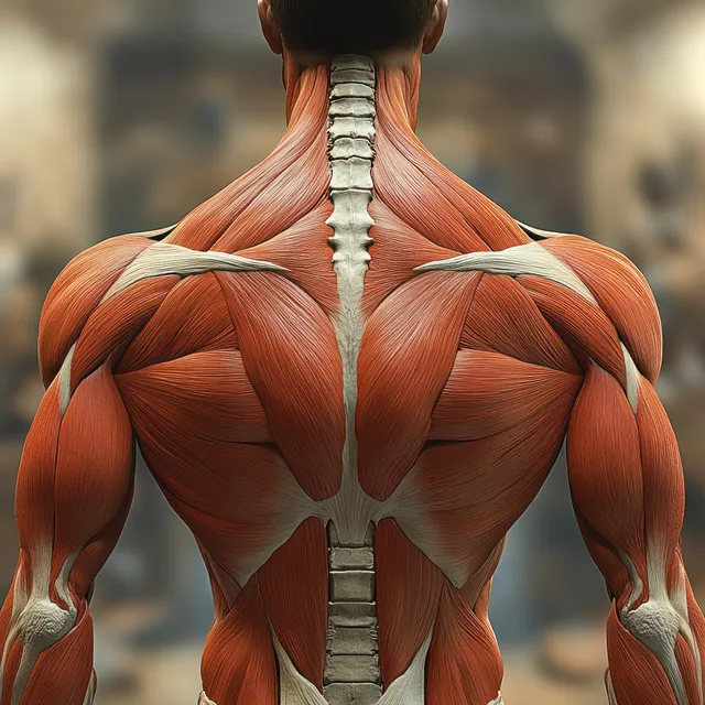
Posterior upper-back anatomy illustration with visible spine and shoulder muscles including rhomboid major and trapezius.
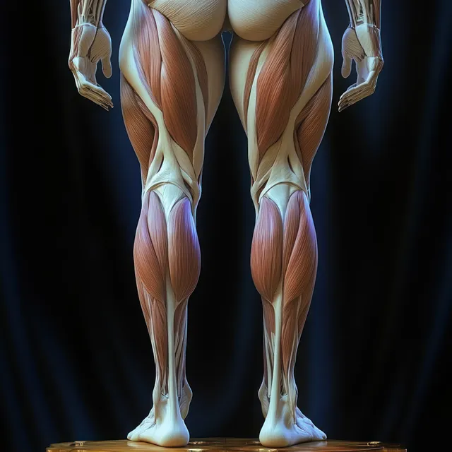
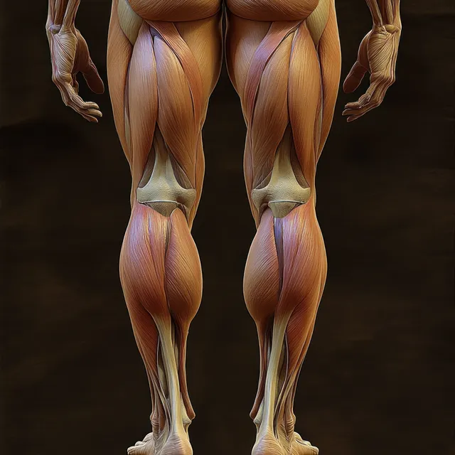
Female lower-body anatomical illustration displaying hip, thigh, and knee muscles with dark lighting and visible tendon lines.
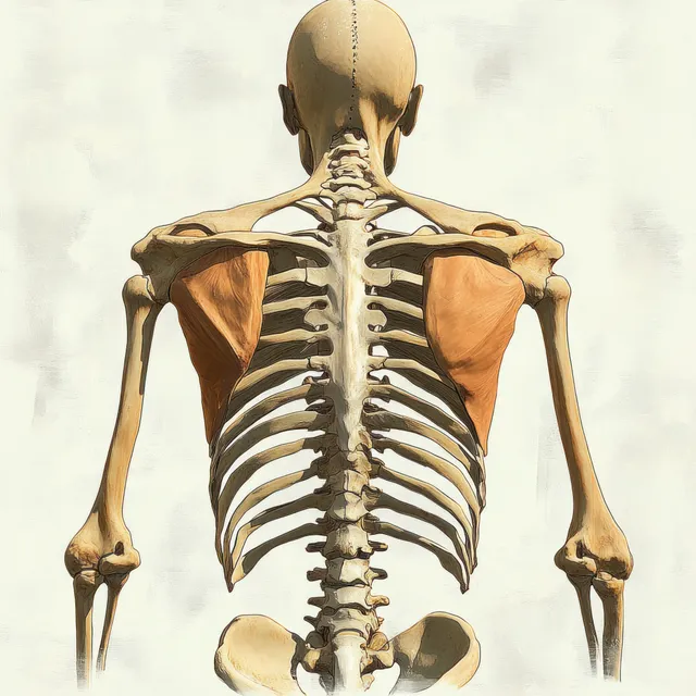
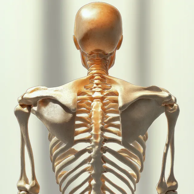
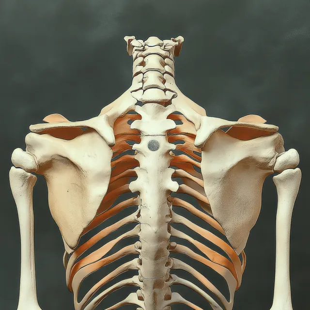
Back view anatomical illustration showing the human skeleton spine and scapulae with ribs and shoulder girdle.
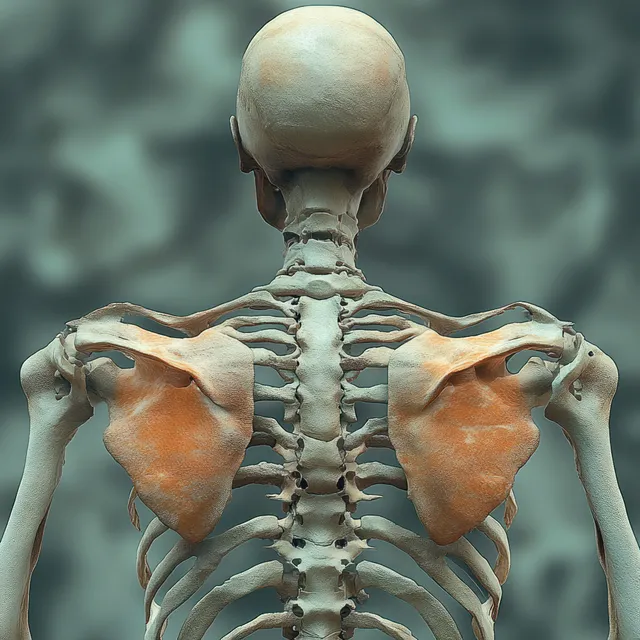
Rear view anatomical illustration of a human skeleton showing spine and shoulder blades with rhomboid minor emphasis.
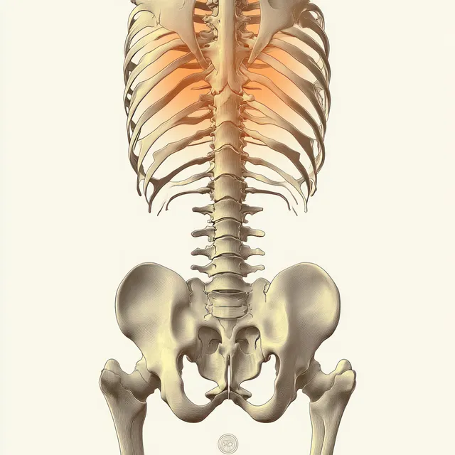
Back view of an anatomical human skeleton illustration with visible vertebrae and pelvic bones against a light background.
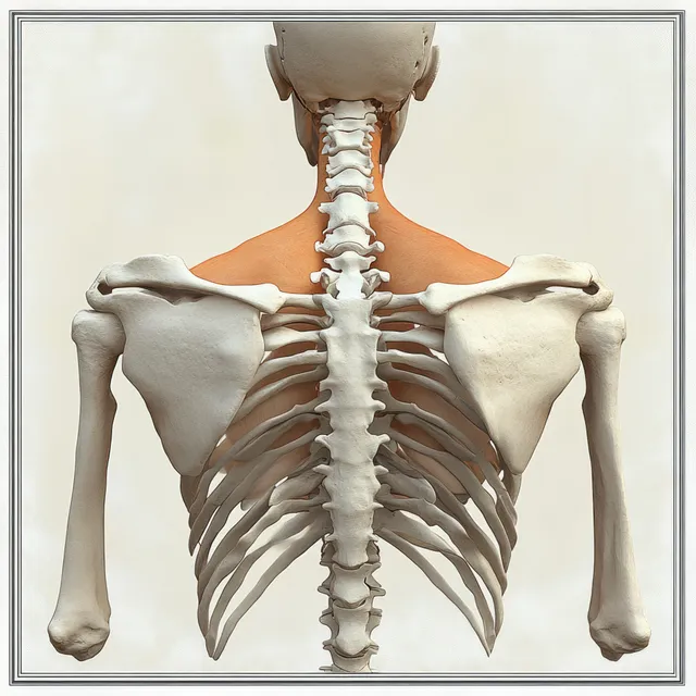
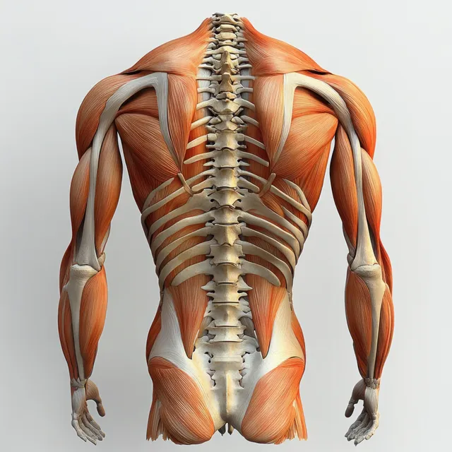
Rear view anatomical illustration displays trapezius and rhomboid muscles along the upper back over the spine.
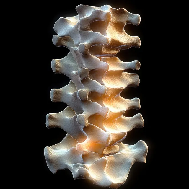
3D anatomical spine model with vertebrae and posterior longitudinal ligament isolated on a black background.
Rear-view human skeleton illustration highlighting the spine and shoulder blades under soft studio light.
Rendered human skeleton viewed from behind with highlighted shoulder joints and supraspinatus muscles.
Posterior view anatomical illustration of the spine and erector spinae muscles in the upper back with detailed vertebrae.
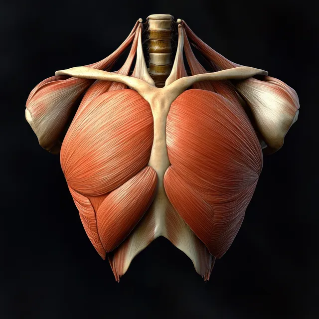
Anterior anatomical illustration of the pectoralis major muscle with ribs, clavicle, and humerus on a black background.
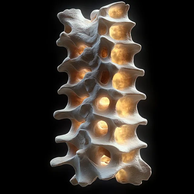
3D vertebral column model with illuminated spinal discs isolated on a dark background.
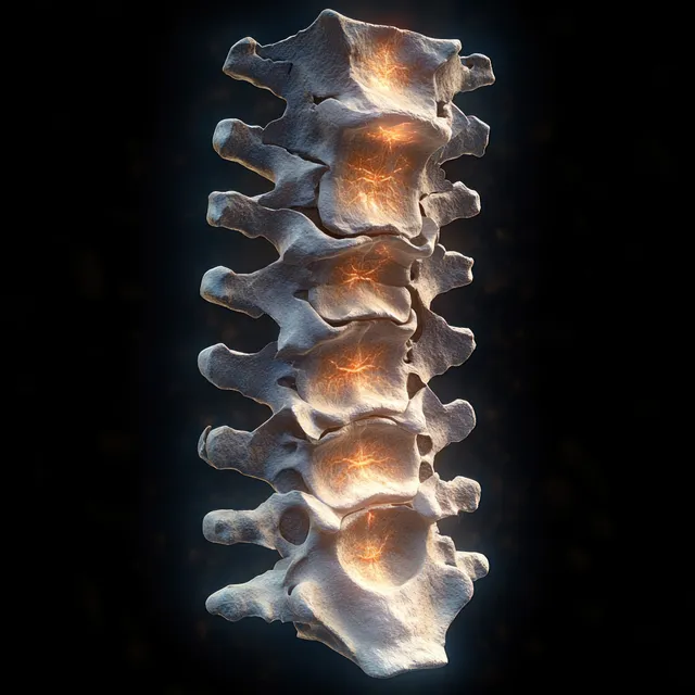
3D render of a human vertebral column with glowing spinal structures on a black background.
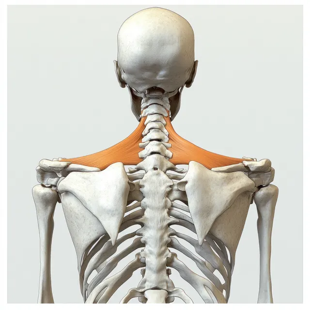
Rear view anatomical illustration reveals the trapezius muscles across the upper back and shoulders surrounding the spine.
Human skeleton model facing forward with visible spine, ribs, and shoulder blades under soft warm studio lighting.
Rear anatomical illustration highlighting female hip and hamstring muscles with visible tendons under low-key lighting.
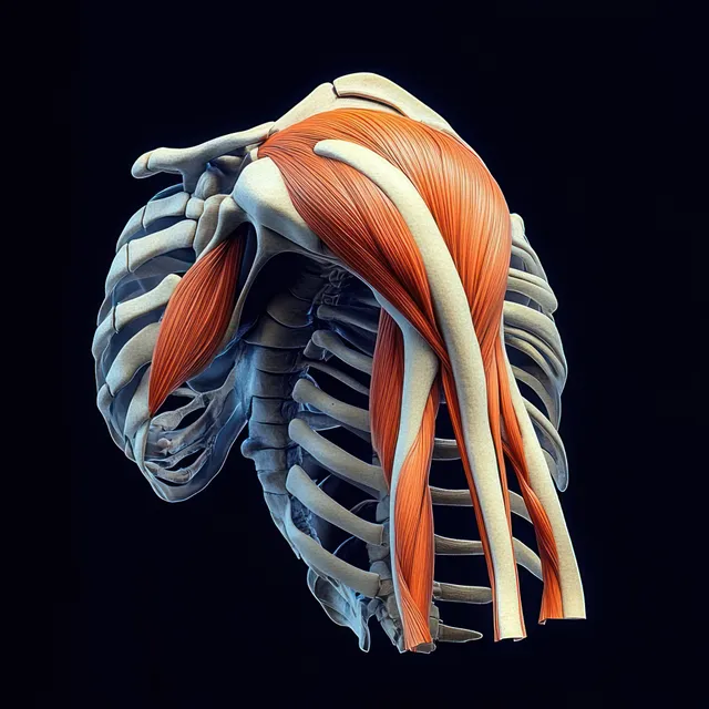
3D medical illustration of shoulder and upper arm muscles and bones with orange muscle bands on a dark background.
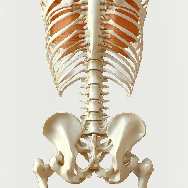
Rear anatomical illustration highlighting erector spinae muscles along the spine with pelvis and lumbar vertebrae detail.
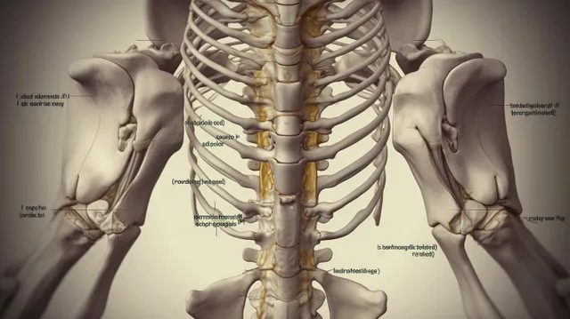
Labeled anatomical illustration of the human pelvis and spine shown in a symmetrical posterior view.
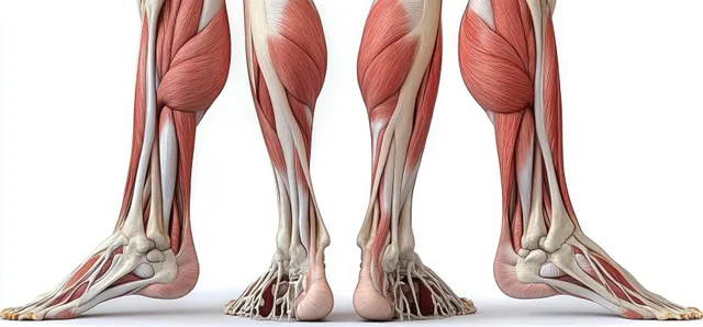
An anatomical cutaway illustration shows leg muscles and bones from front and back views on a white background.
Rear-view human skeleton model focusing on shoulder joints with dramatic chiaroscuro lighting.
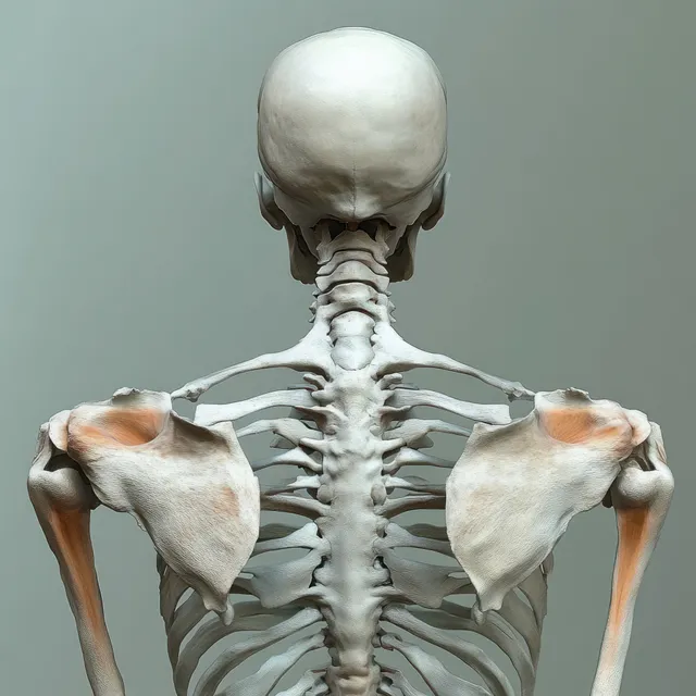
Rear view human skeleton model focusing on the spine, shoulder blades, and ribcage.
Rear view human skeleton illustration displaying the spine, sacrum, and hip pelvis bones on a light background.
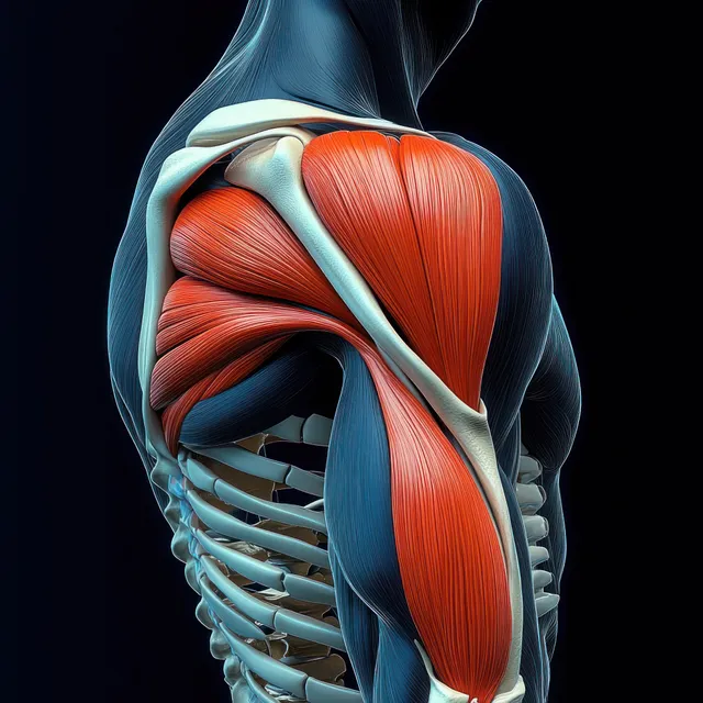
3D anatomical illustration of the highlighted deltoid and shoulder muscles over a skeletal torso on a dark background.
Posterior Anatomical View images | StockPico