Scientific Education images

Explore stock images tagged scientific education
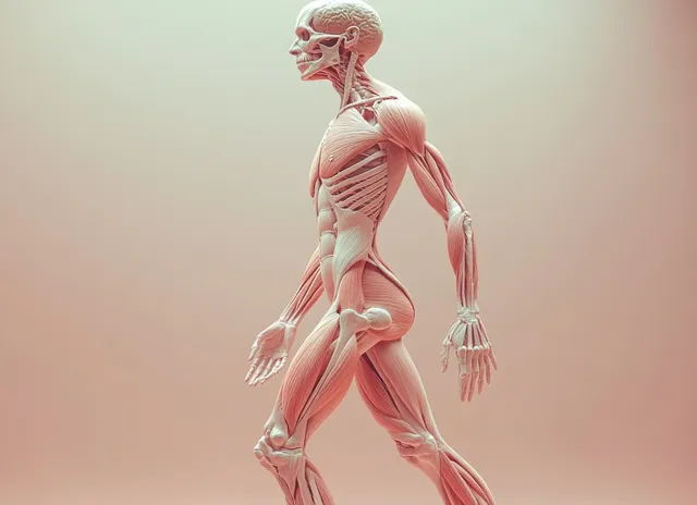
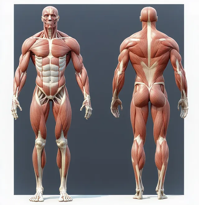
3D human figure anatomy view with transparent muscles and skeleton, showing labeled blood vessels on a soft background.
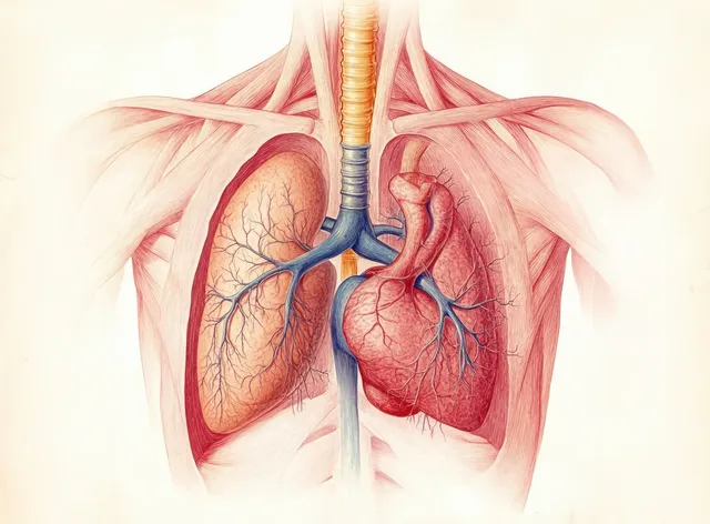
Illustrated human torso showing lungs and heart inside a rib cage with branching bronchial and blood vessels.
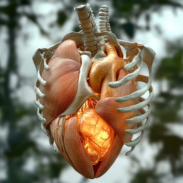
A suspended anatomical view of the heart and ribcage with glowing orange muscle tissue against a blurred background.
Connected transparent blue spheres form a molecular chain with glowing bubbles inside on a soft light blue background.
Glossy atom spheres linked by molecular bonds float over a blurred laboratory and biotech research backdrop.
Two-view anatomical illustration of a male body showing muscles, skeleton, and internal organs with dramatic lighting on a dark background.
A hand holds a pen pointing at the rib cage and spine on a detailed anatomical model under warm light.
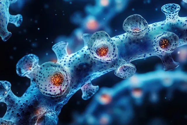
Blue translucent virus-like cells with bulbous protrusions and orange glowing cores float in a dark blurred background.
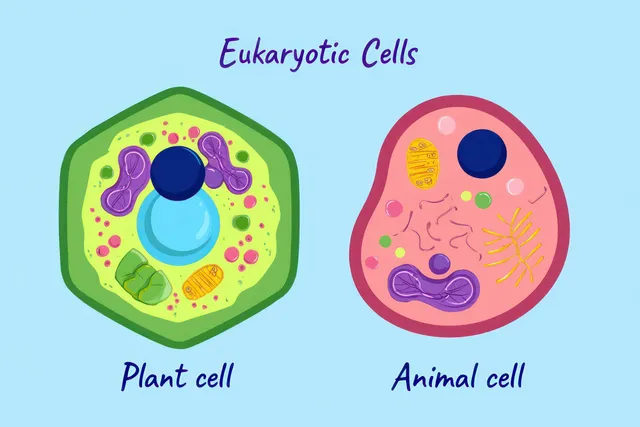
Two eukaryotic cell diagrams, plant and animal, display organelles like nucleus, chloroplasts, and mitochondria on a blue background.
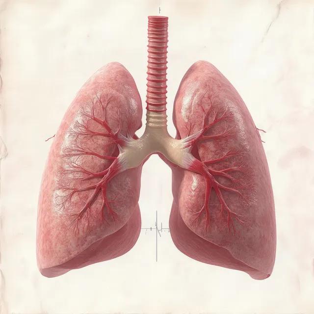
Illustrated human lungs with a central trachea and branching bronchi on a pale light background.
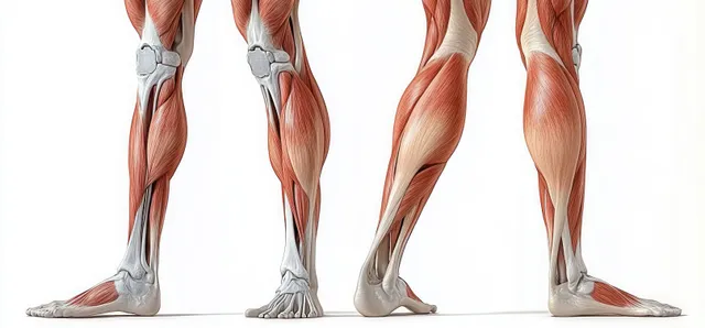
Anatomical leg study displaying muscles and bones in front and back positions against a white background.
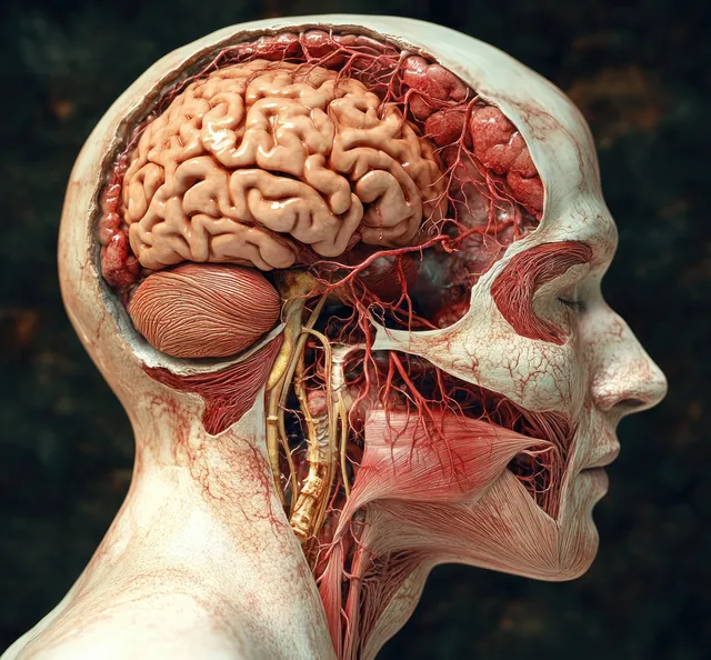
Anatomical cutaway of a human skull revealing the brain cross-section and nervous system pathways.
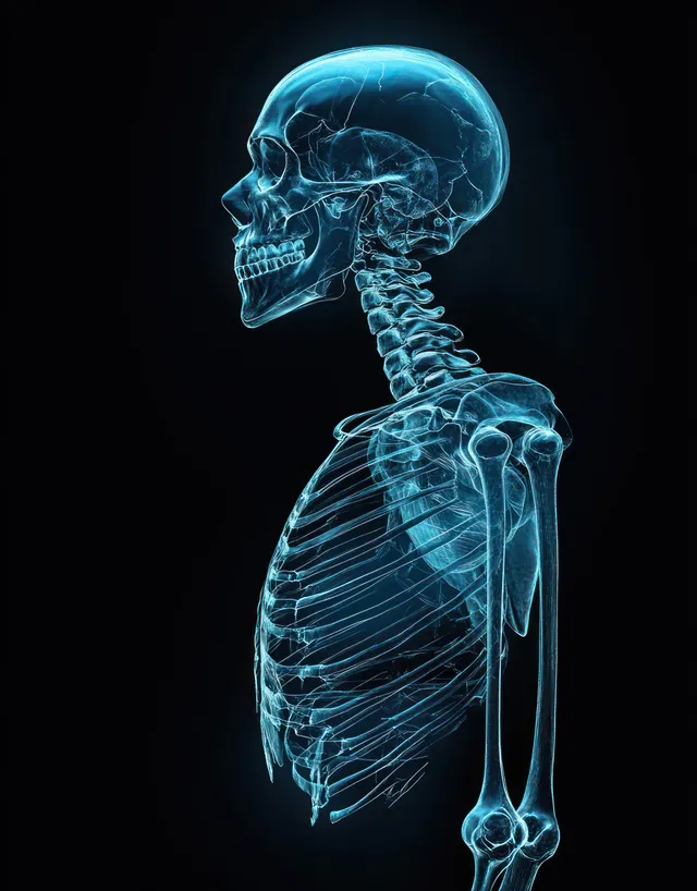
Glow-lit blue male skeleton in three-quarter view with skull, spine, ribs, and joints on a dark background.
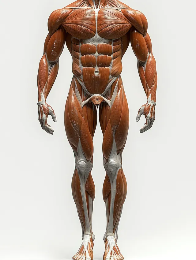
Standing male figure showing detailed leg muscle groups, tendons, knee, ankle, and foot contours on a plain background.
Glowing red blood cells flowing along a dark vein stream with scattered microscopic particles around them.
Glowing 3D atoms and bonded molecular shapes float within a misty abstract science background.
A floating molecular cluster of glowing atom spheres hangs against a soft blue blurred laboratory background.
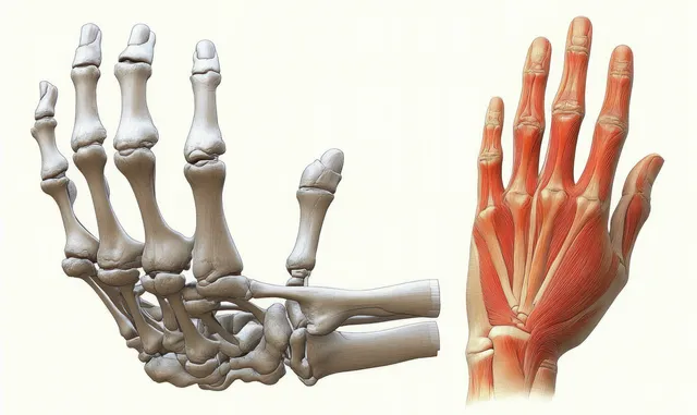
An anatomical illustration shows a human hand with bones, muscles, and tendons in dorsal view.
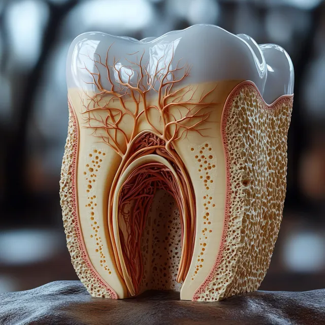
A human molar cross-section displays enamel, dentin, a pulp chamber with nerves and vessels, and porous root bone texture.
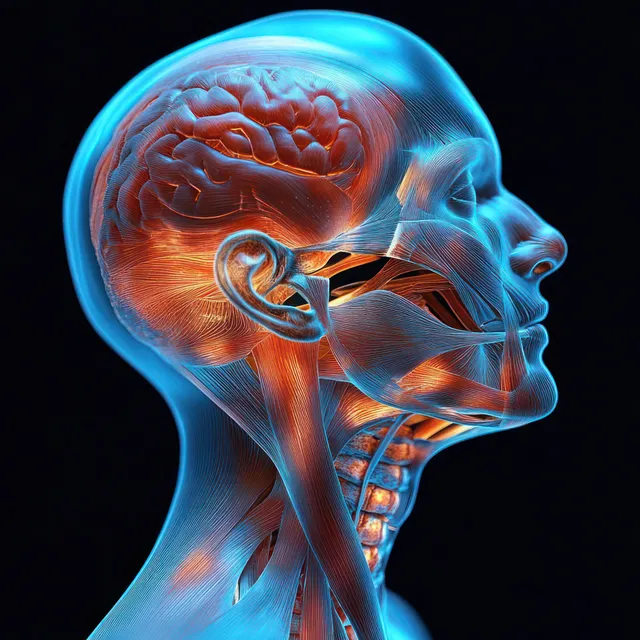
Profile view anatomical illustration of the human neck with highlighted muscles, nerves, and connective tissues.
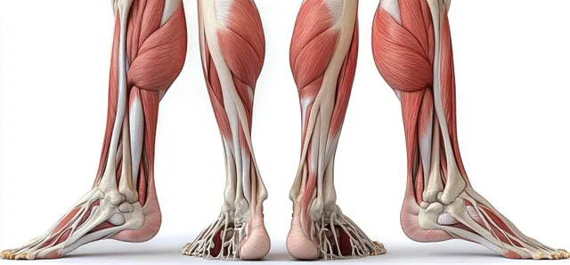
An anatomical cutaway illustration shows leg muscles and bones from front and back views on a white background.
A masked female scientist in protective gear peers into a microscope with a conical flask on a lab bench.
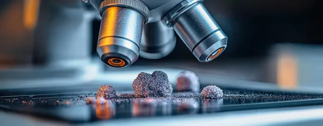
Microscope examining textured samples on a slide in a laboratory.
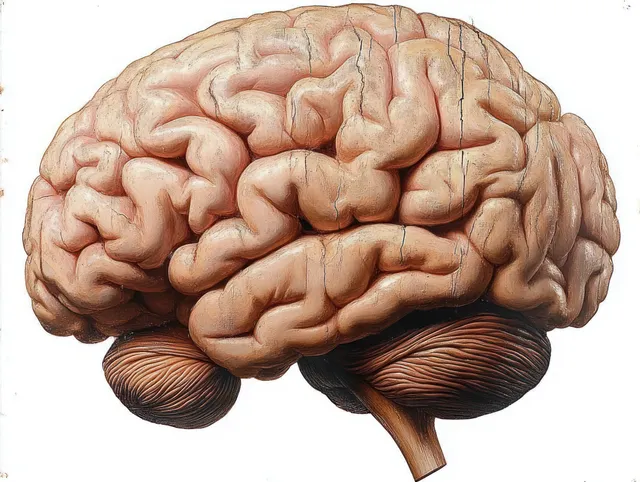
Human brain illustration highlighting cerebral cortex folds, cerebellum, and brainstem with detailed shading.