Sports Injury images

Explore stock images tagged sports injury

Physiotherapist massaging a patient’s knee during a rehabilitation appointment in a clinic.
3D render of a human skeleton with glowing lumbar vertebrae against a blue, bokeh-lit background.
Physiotherapist’s hands massage an athlete’s sore knee next to a doctor holding a stethoscope.
Runner holding her aching knee on an asphalt road with a glowing pain spot near the joint.
Side-profile head and neck medical illustration with neon glowing lines highlighting the scapula and shoulder.
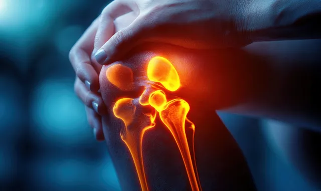
Hand gripping a painful knee with glowing bones overlay suggesting inflamed internal joint.
Physiotherapist massages an athlete’s knee while examining the injured leg in a clinic.
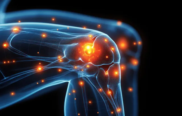
A glowing human knee joint is shown with inflammation points on a dark background.
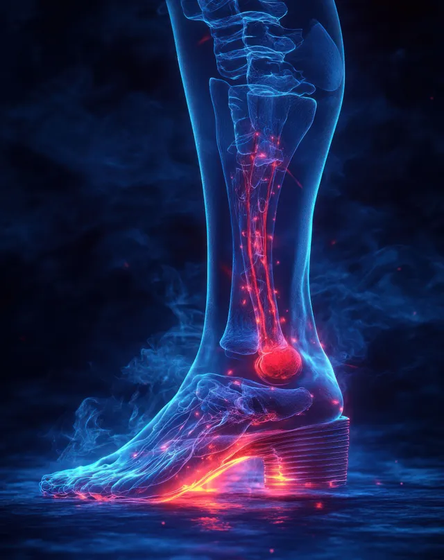
3D ankle joint X-ray illustration with red inflammation highlights on a dark blue medical background.
3D human skeleton spine illustration highlights a glowing herniated lumbar disc with red and blue pain signals.
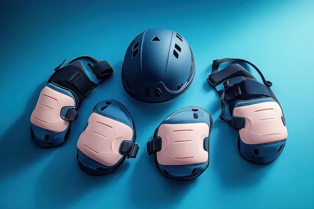
Blue helmet and pink knee, elbow, and wrist pads are neatly arranged on a turquoise background.
A therapist’s hands massage the athlete’s back on a padded table in a softly lit clinic.
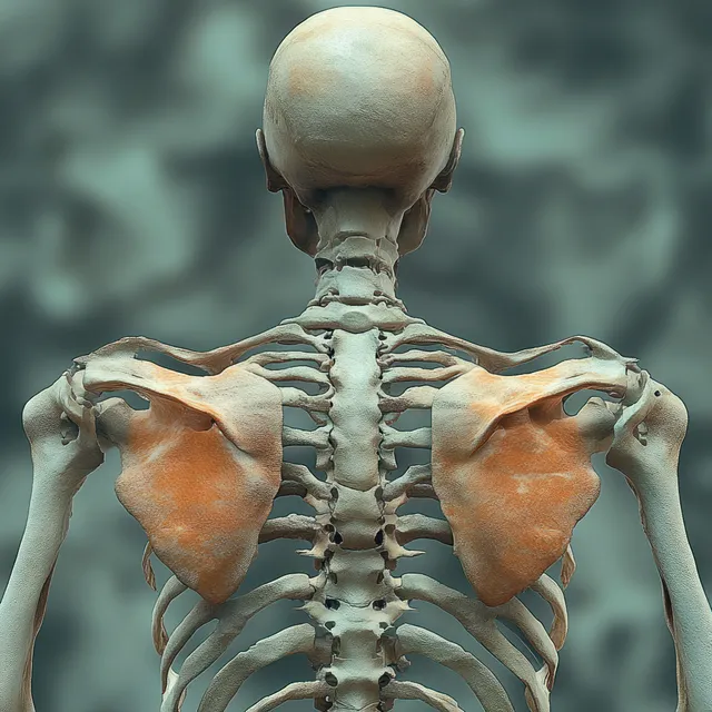
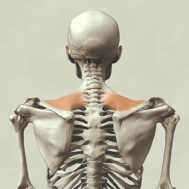
Rendered human skeleton viewed from behind with highlighted shoulder joints and supraspinatus muscles.
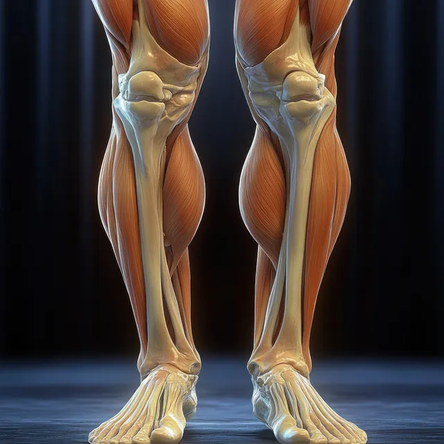
Anterior view 3D anatomical legs highlighting quadriceps muscles and knee joint with visible bone contours.
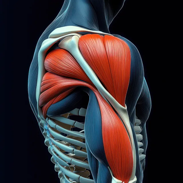
3D anatomical illustration of the highlighted deltoid and shoulder muscles over a skeletal torso on a dark background.
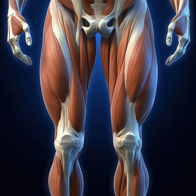
Male leg anatomy illustration highlighting quadriceps muscles with visible femur and tendons under blue dramatic lighting.
Back view of a human skeleton with highlighted shoulder joints and supraspinatus muscle near the scapula.
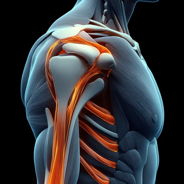
Anatomical shoulder and triceps view showing humerus, scapula, tendons, and glowing orange highlights on a dark background.
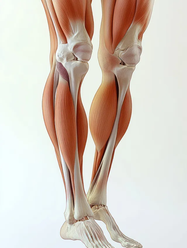
3D anatomy illustration showing hamstring muscles on both legs from a lateral view around the knee.
3D render of anterior knee anatomy showing femur, patella, meniscus, cartilage, and surrounding ligaments on a dark background.
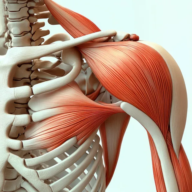
Anterior shoulder anatomy model showing highlighted deltoid muscle with visible clavicle, scapula, and rib cage.
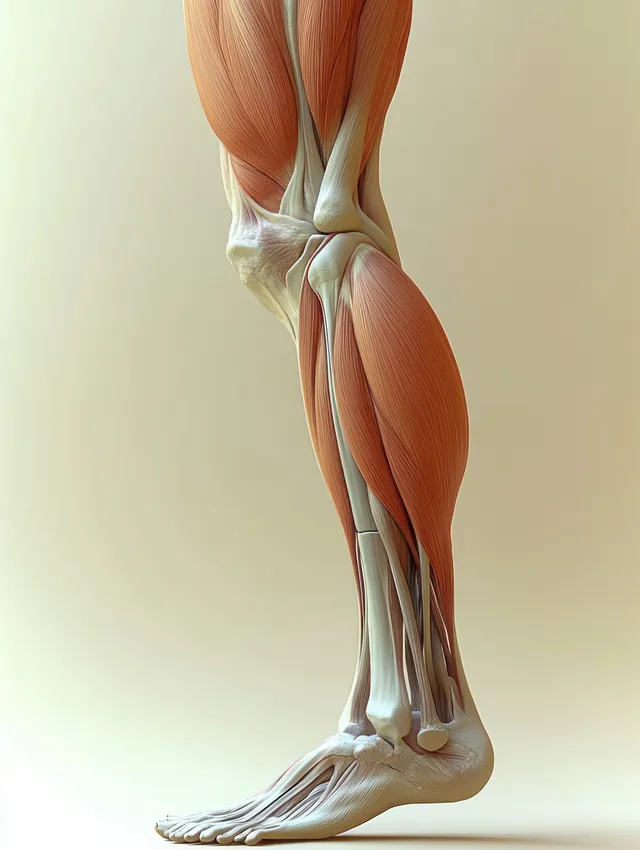
Lateral view anatomical illustration of the hamstring muscles and knee joint with highlighted thigh tendons.
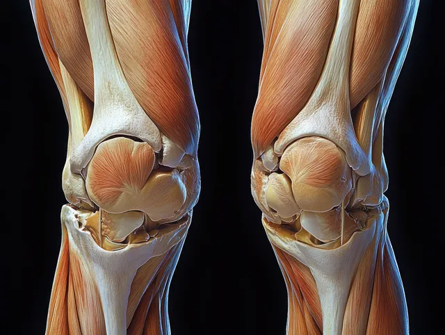
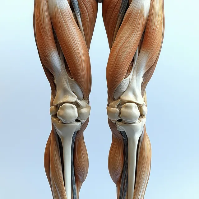
Anatomical illustration of both knees and thighs with visible quadriceps muscles, bones, and knee joint alignment against a plain background.
3D render of a human knee joint with a glowing red inflamed patella area on blue bones.
Sports Injury images | StockPico