StockPico
免费 4K 图库,为现代创意团队打造
首页
分类
搜索
个人中心
帮助
🇨🇳 ZH
▾
Health
/
Image details
Health
Asset ID YE5BBAF9C44Q06EF
standard
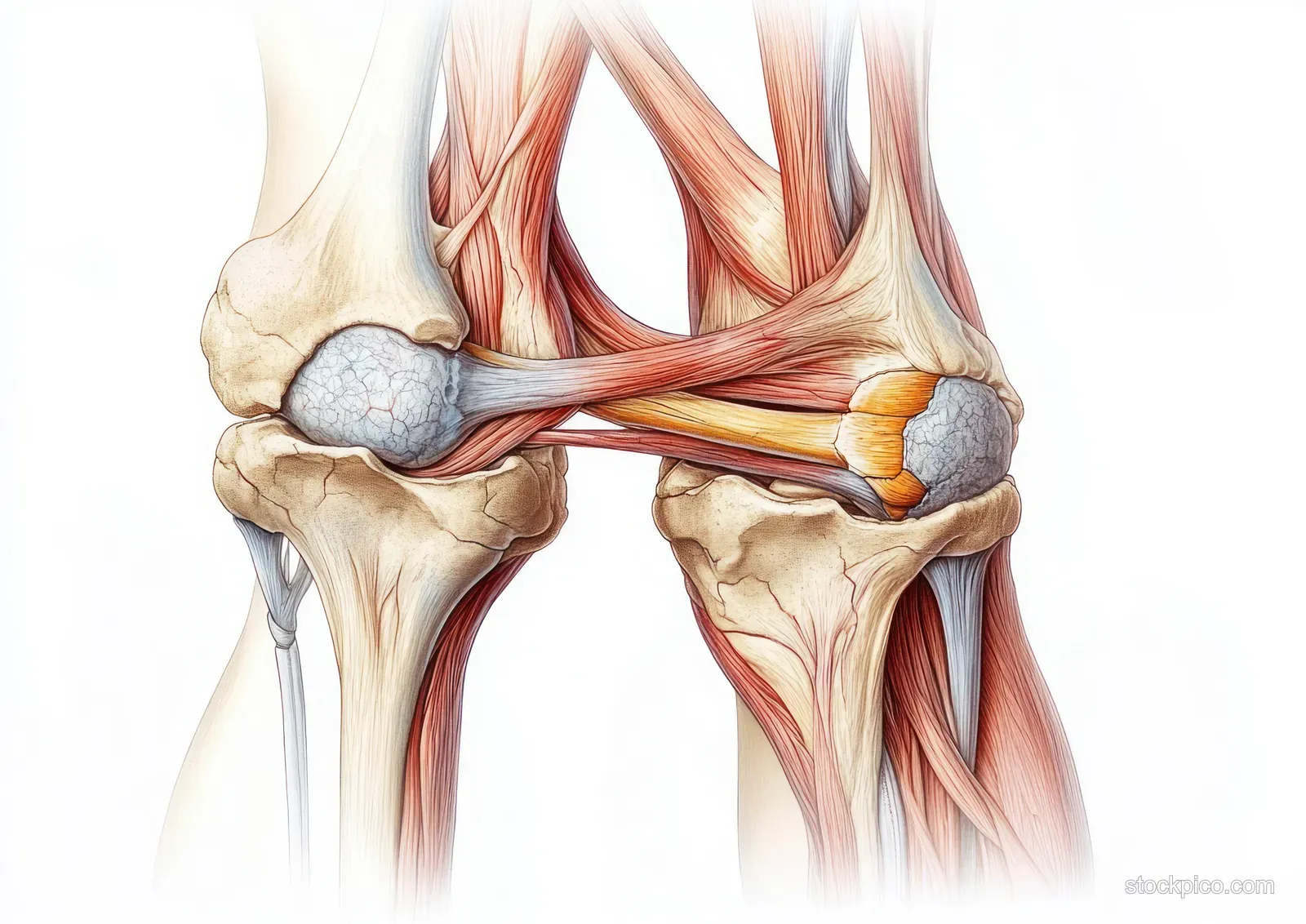
膝关节横截面解剖插图,展示股骨、髌骨、半月板和肌腱
一幅用于教学的膝关节横截面医学图,突出显示髌骨和股骨周围的肌肉、韧带、软骨和半月板。
Color palette
Keywords
解剖学
人体
膝关节
解剖学图表
横截面
股骨
Show more
Tags
解剖学插图
横截面示意图
医疗可视化
教育类科学
详细的组织渲染
柔和的漫射灯光
Popular searches:
Business meeting stock images
·
Office background stock images
·
Medical team stock images
·
Technology background stock images
·
Real estate house stock images
·
Healthy food stock images
相关编辑图片素材