Connective Tissue images

Explore stock images tagged connective tissue
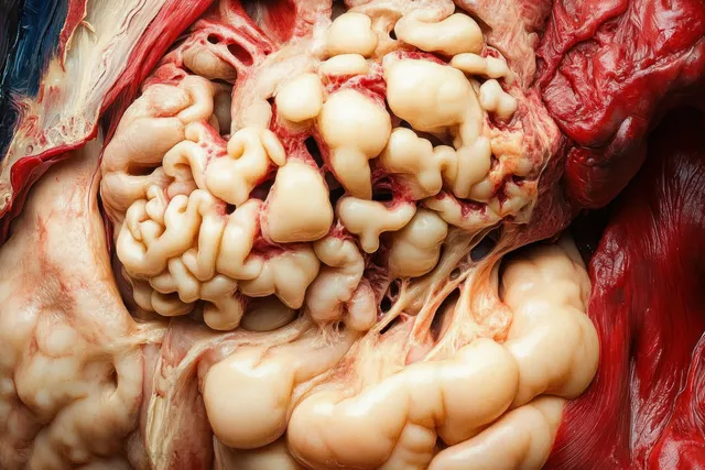
Macro close-up of animal internal organs with pale fat deposits, red muscle tissue, and layered connective structures.
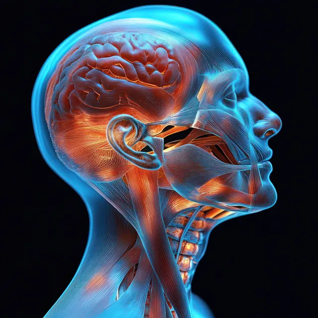
Side profile anatomical illustration of human neck muscles, tendons, and connective tissues with blue and orange glow.
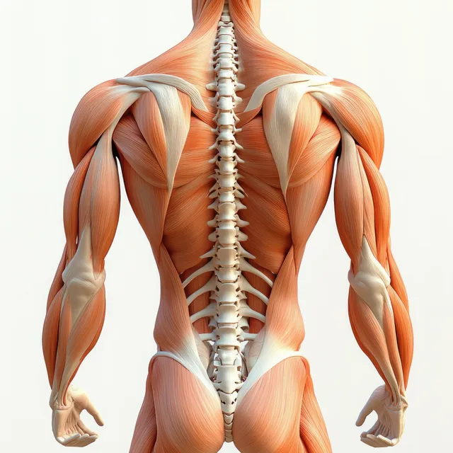
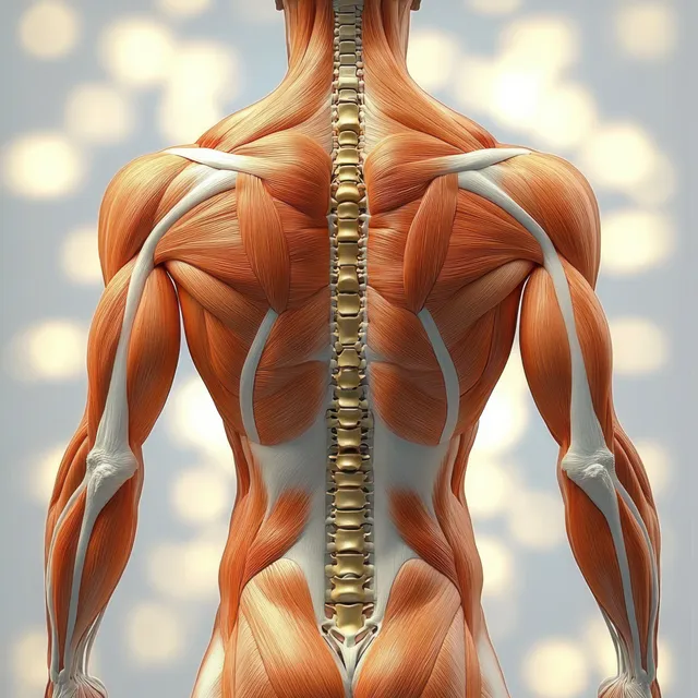
Posterior view anatomical illustration of the spine and erector spinae muscles in the upper back with detailed vertebrae.
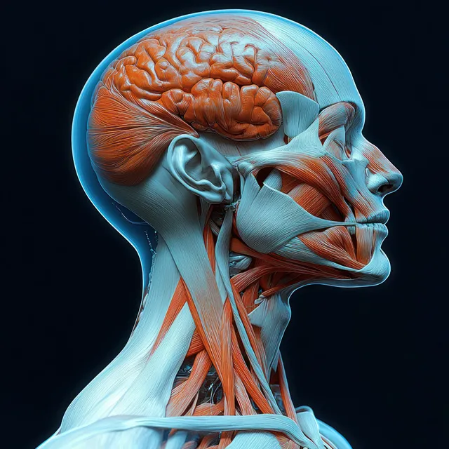
Profile view anatomical illustration of the human neck with highlighted muscles, nerves, and connective tissues.
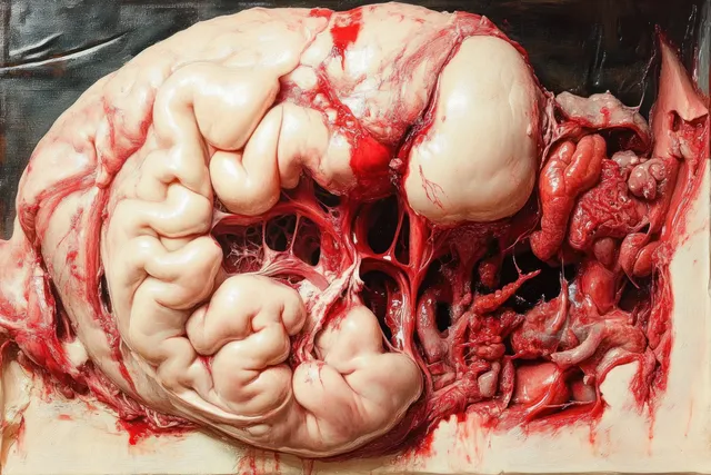
An anatomical cross-section displays pale internal organs with intricate red blood vessels and textured tissue.
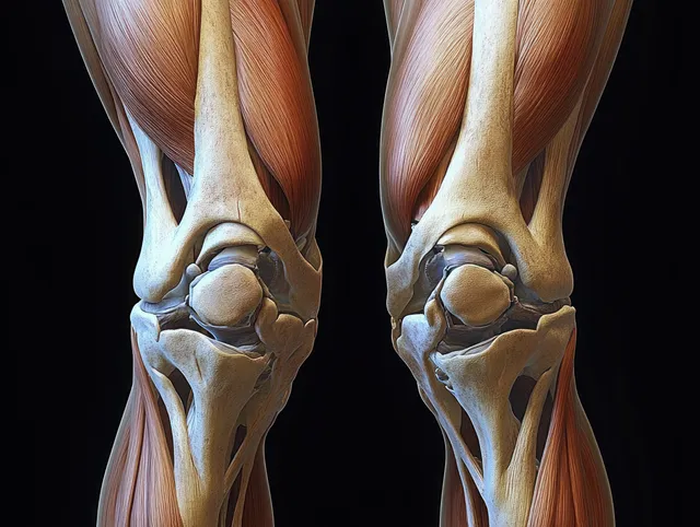
Symmetrical anatomical illustration of both knees showing femur and tibia with cartilage, meniscus, tendons, and ligaments on a dark background.
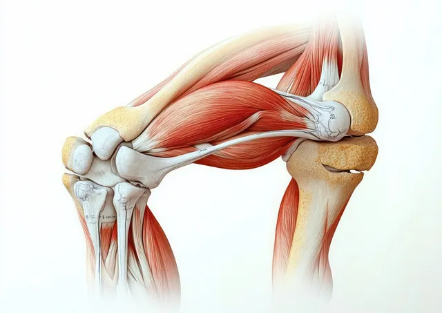
3D anatomical illustration of the human knee joint with exposed muscles, tendons, ligaments, and kneecap against a light background.
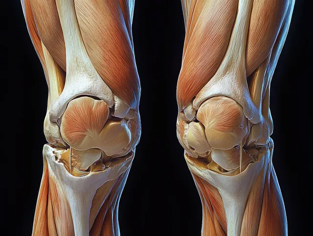
3D render of anterior knee anatomy showing femur, patella, meniscus, cartilage, and surrounding ligaments on a dark background.
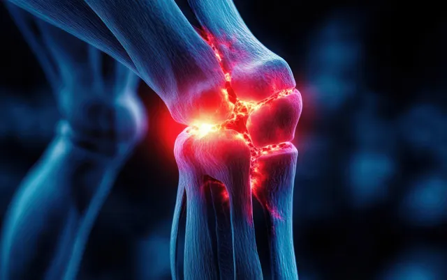
Digital knee joint model with glowing red inflamed areas around the joint and surrounding bones.
Rear-view anatomical illustration reveals the erector spinae muscles along the upper spine with detailed vertebrae.
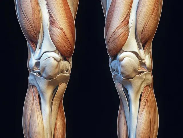
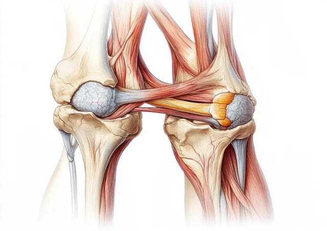
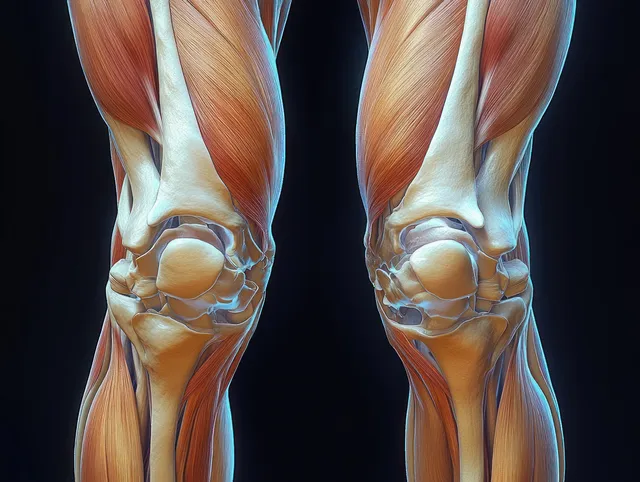
Detailed illustration of human knees showing cartilage, meniscus, ligaments, and surrounding muscles in two mirrored views.
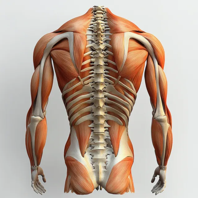
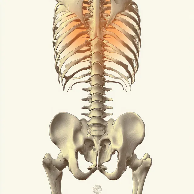
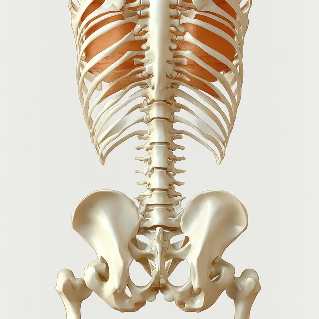
Rear anatomical illustration highlighting erector spinae muscles along the spine with pelvis and lumbar vertebrae detail.
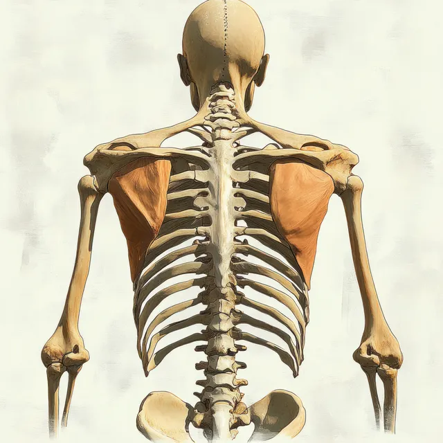
Back view anatomical illustration showing the human skeleton spine and scapulae with ribs and shoulder girdle.
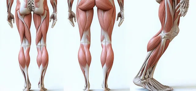
Front and back views of a male leg anatomy illustration with highlighted muscles, bones, tendons, and ligaments on a light background.
An anatomical cross-section illustration of the human knee showing patella, meniscus, cartilage, ligaments, and tendons.
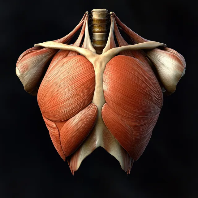
Anterior anatomical illustration of the pectoralis major muscle with ribs, clavicle, and humerus on a black background.
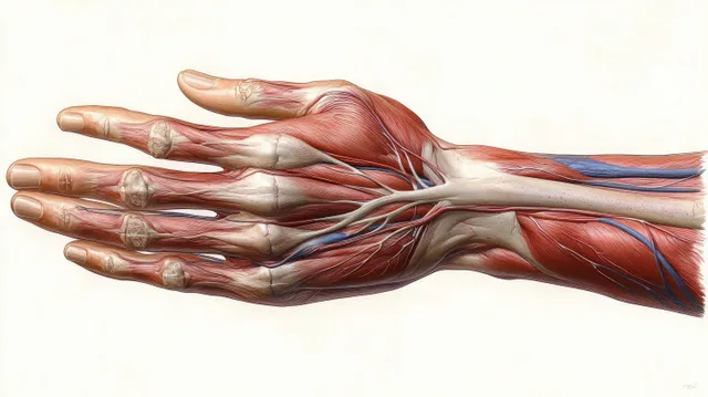
Anatomical illustration of a female hand ventral view with visible muscles, tendons, arteries, and veins.
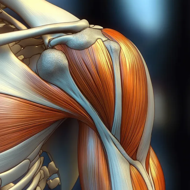
Anatomical illustration of shoulder joint muscles and bones with deltoid and triceps on a white background.
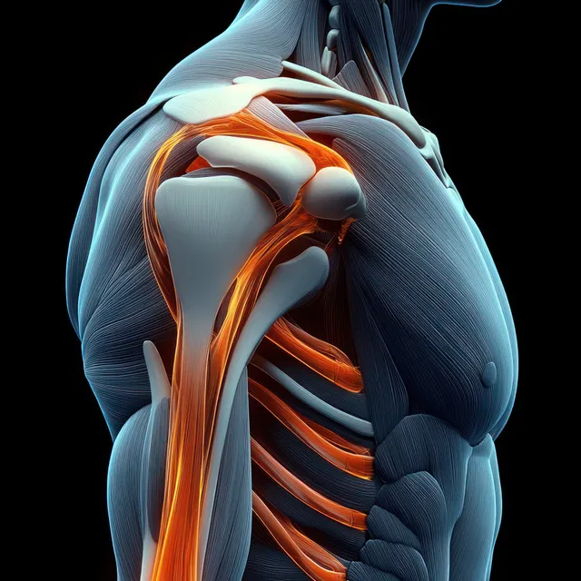
Anatomical shoulder and triceps view showing humerus, scapula, tendons, and glowing orange highlights on a dark background.
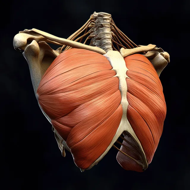
Red pectoral muscles are highlighted on an anterior skeletal torso against a dark background.
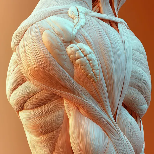
Renaissance anatomy illustration highlights deltoid and triceps muscles with visible tendons and soft colored shading.
Rear view human skeleton illustration displaying the spine, sacrum, and hip pelvis bones on a light background.
Back view of an anatomical human skeleton illustration with visible vertebrae and pelvic bones against a light background.
Front anatomical view of human knees displaying femur, tibia, ligaments, tendons, and cartilage in vivid detail.