Knee Anatomy images

Explore stock images tagged knee anatomy
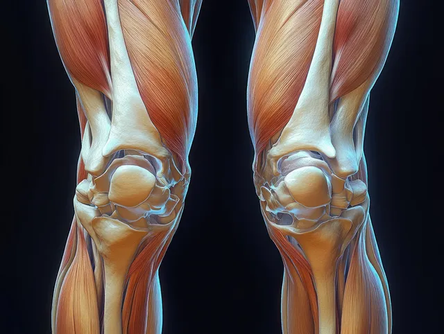
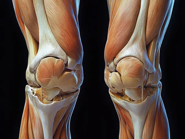
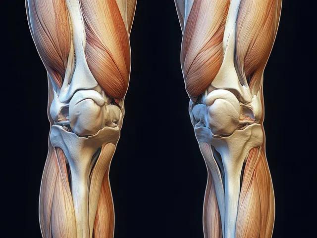
Front anatomical view of human knees displaying femur, tibia, ligaments, tendons, and cartilage in vivid detail.
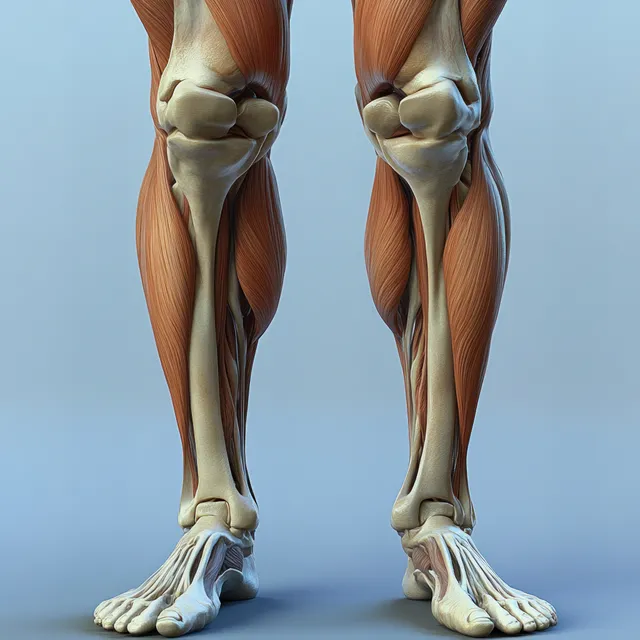
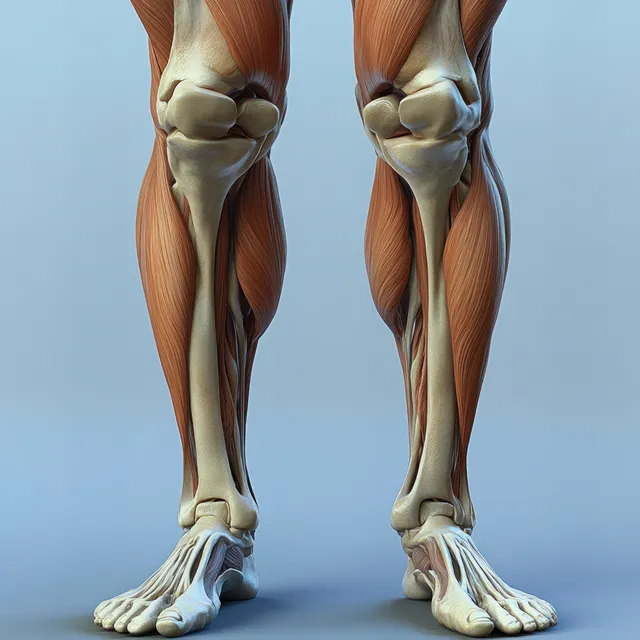
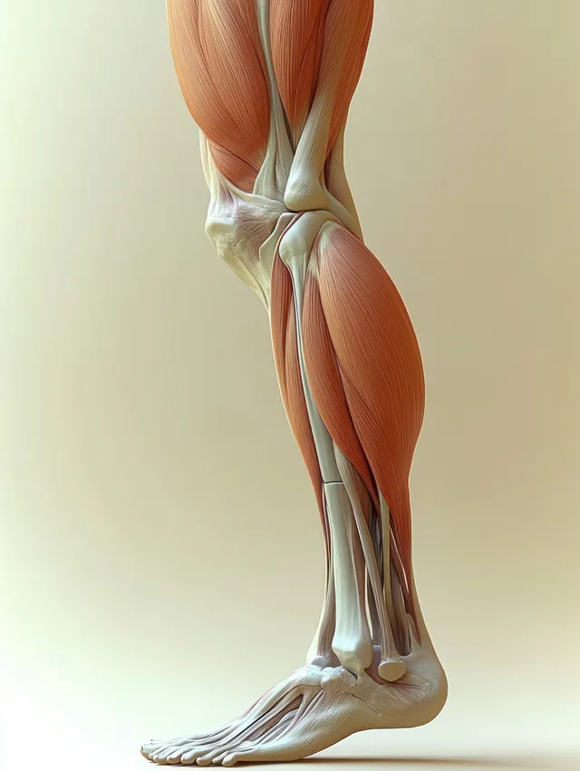
Illustrated legs show quadriceps muscles and detailed knee joints with exposed bone structure in a clinical setup.
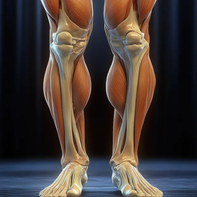
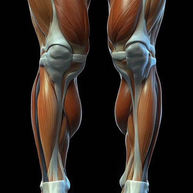
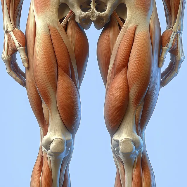
Anterior view 3D anatomical legs highlighting quadriceps muscles and knee joint with visible bone contours.
3D render of anterior knee anatomy showing femur, patella, meniscus, cartilage, and surrounding ligaments on a dark background.
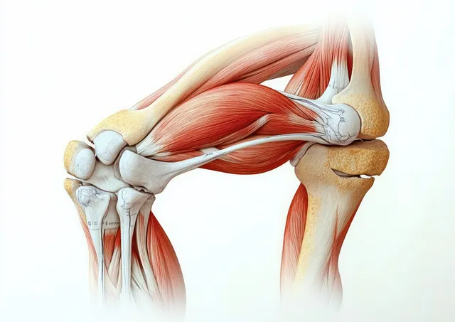
3D anatomical illustration of the human knee joint with exposed muscles, tendons, ligaments, and kneecap against a light background.
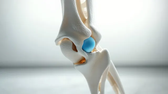
3D knee joint model with highlighted bones and cartilage resting on a smooth blue surface.
Lateral view anatomical illustration of the hamstring muscles and knee joint with highlighted thigh tendons.
Anatomical illustration showing quadriceps and hamstrings muscles on both legs with highlighted knee joints.
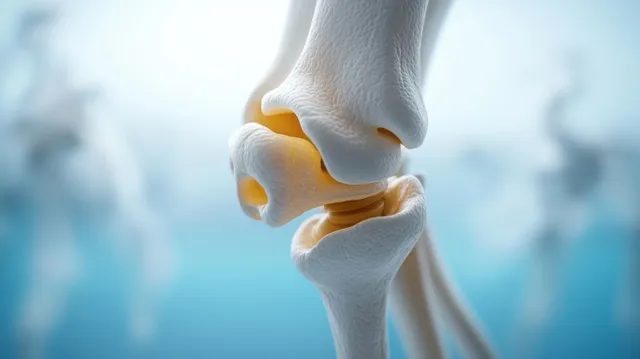
3D knee joint anatomy close-up with highlighted cartilage and meniscus layers on a pale background.
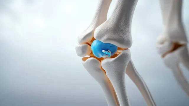
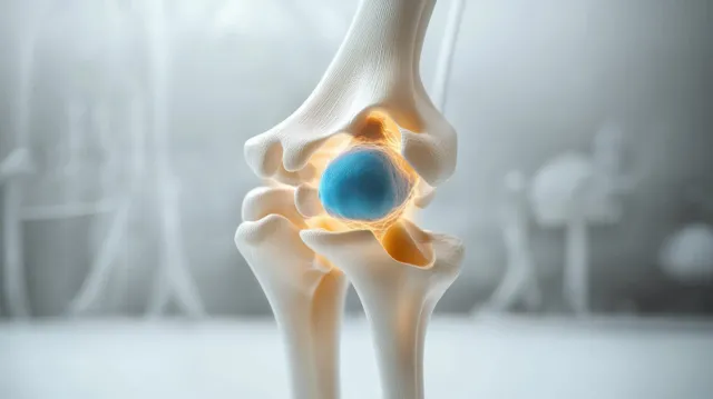
Close-up view of a knee joint anatomy model with blue meniscus and textured cartilage under soft lighting.
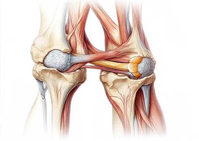
Detailed illustration of human knees showing cartilage, meniscus, ligaments, and surrounding muscles in two mirrored views.
Orange glowing knee joint anatomy illustration with a hand resting on the thigh over a dark background.
3D anatomical knee joint model with highlighted blue meniscus and cartilage on a softly blurred background.
3D knee joint rendering with highlighted cartilage and diffused soft blue background lighting.
3D render of a human knee joint with a glowing red inflamed patella area on blue bones.
Digital knee joint model with glowing red inflamed areas around the joint and surrounding bones.
Hyper-realistic view of male thighs and calves emphasizing quadriceps muscle groups and knee joints under blue backlight.
A hand holds a knee as glowing internal bones show beneath the skin on a blue background.
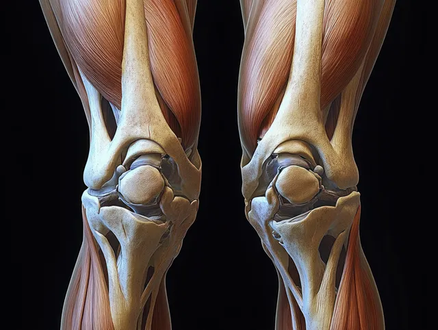
Symmetrical anatomical illustration of both knees showing femur and tibia with cartilage, meniscus, tendons, and ligaments on a dark background.
Neon 3D knee joint illustration with blue and red glowing pain zones on a dark background.
3D knee joint anatomy with femur and tibia, highlighting an inflamed pain area using a bright orange glow.
An anatomical cross-section illustration of the human knee showing patella, meniscus, cartilage, ligaments, and tendons.
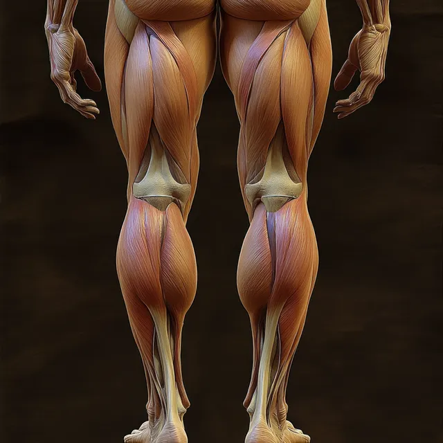
Rear anatomical illustration highlighting female hip and hamstring muscles with visible tendons under low-key lighting.
Knee Anatomy images | StockPico