Ligaments images

Explore stock images tagged ligaments
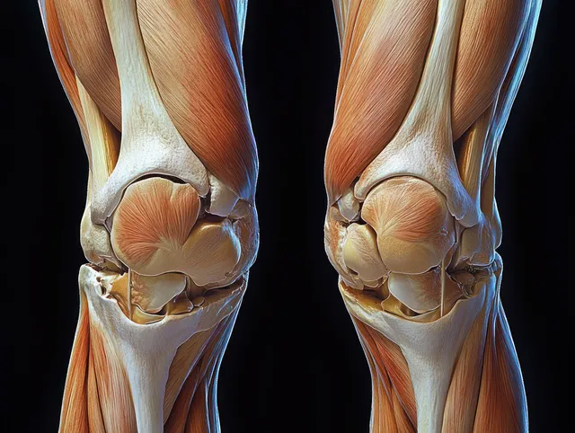
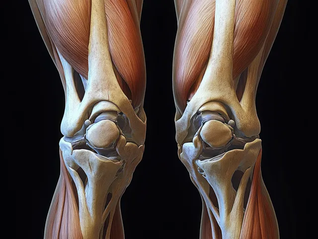
Front anatomical view of human knees displaying femur, tibia, ligaments, tendons, and cartilage in vivid detail.
3D render of anterior knee anatomy showing femur, patella, meniscus, cartilage, and surrounding ligaments on a dark background.
Labeled shoulder joint anatomy illustration showing clavicle, scapula, humerus, and ligaments on a white background.
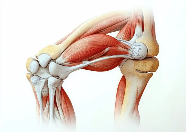
3D anatomical illustration of the human knee joint with exposed muscles, tendons, ligaments, and kneecap against a light background.
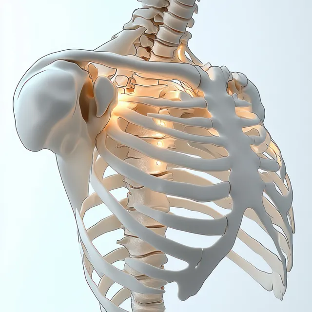
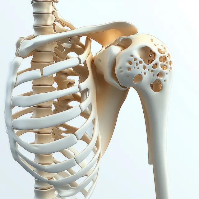
3D anatomical shoulder joint view with scapula and humerus bones and ligaments on a white background.
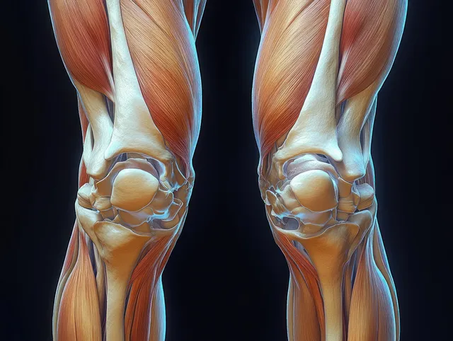
Symmetrical anatomical illustration of both knees showing femur and tibia with cartilage, meniscus, tendons, and ligaments on a dark background.
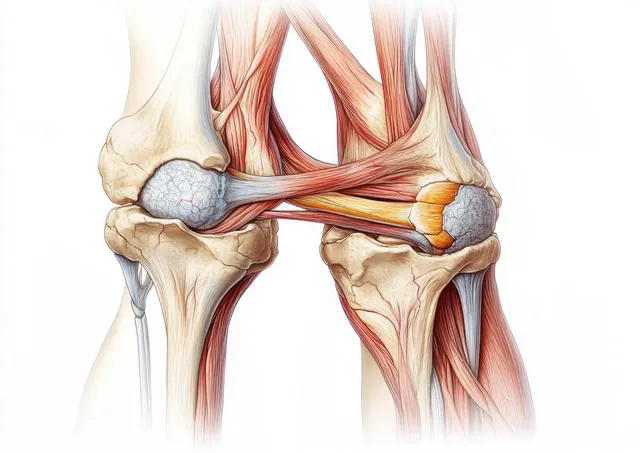
An anatomical cross-section illustration of the human knee showing patella, meniscus, cartilage, ligaments, and tendons.
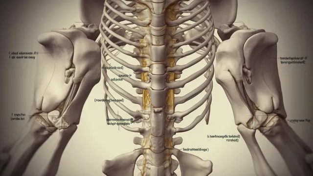
Labeled anatomical illustration of the human pelvis and spine shown in a symmetrical posterior view.
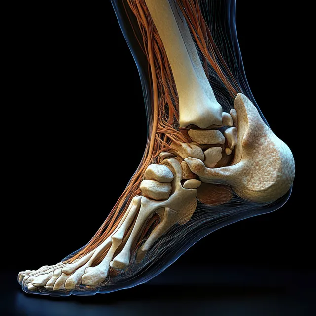
Medial x-ray style view of a human foot showing heel bones, ankle joints, and glowing tendons on a dark background.
Hands hold a human knee as an orange glowing area marks inflammation or pain on the joint.
Hand holding an aching knee with a glowing knee joint and inflammation effect.
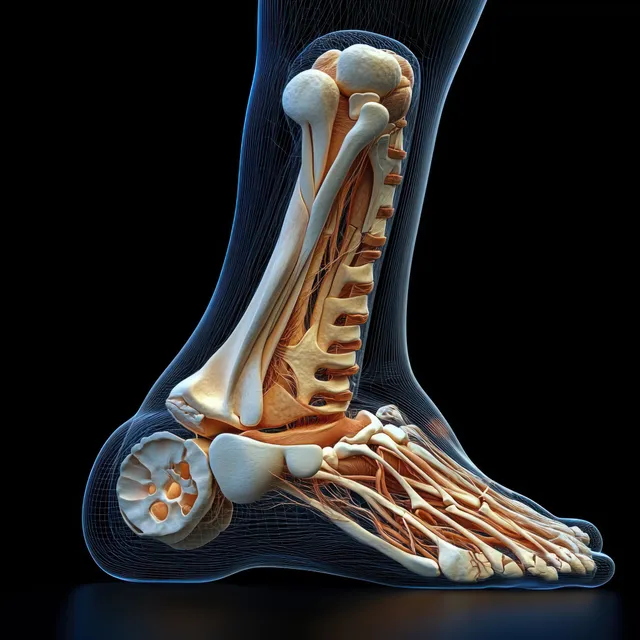
Surreal anatomical illustration of a human foot with exposed bones and tendons under dramatic rim lighting.
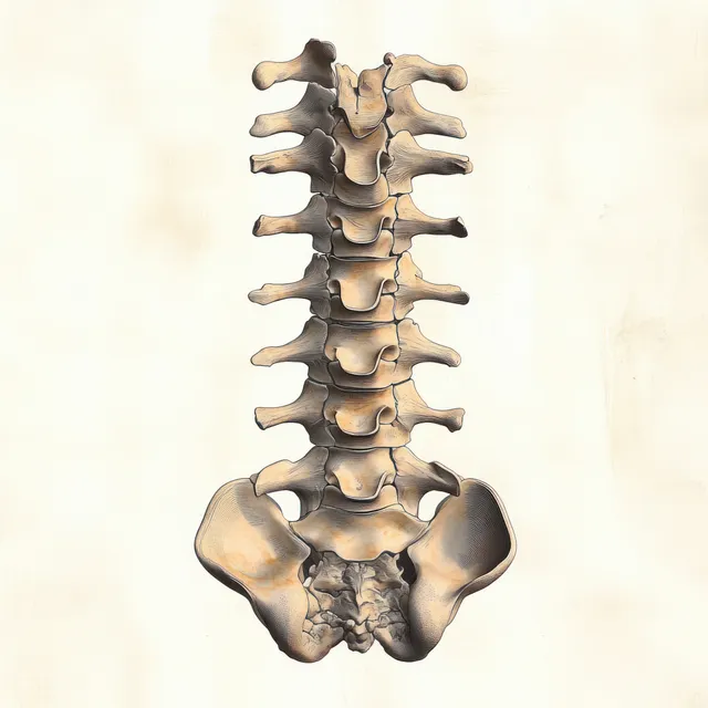
An anatomical illustration of the human spine and lumbar vertebrae with the posterior longitudinal ligament highlighted.
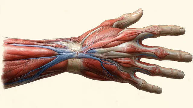
Ventral anatomical illustration of a female hand with visible muscles, arteries, veins, and tendons laid over bones.
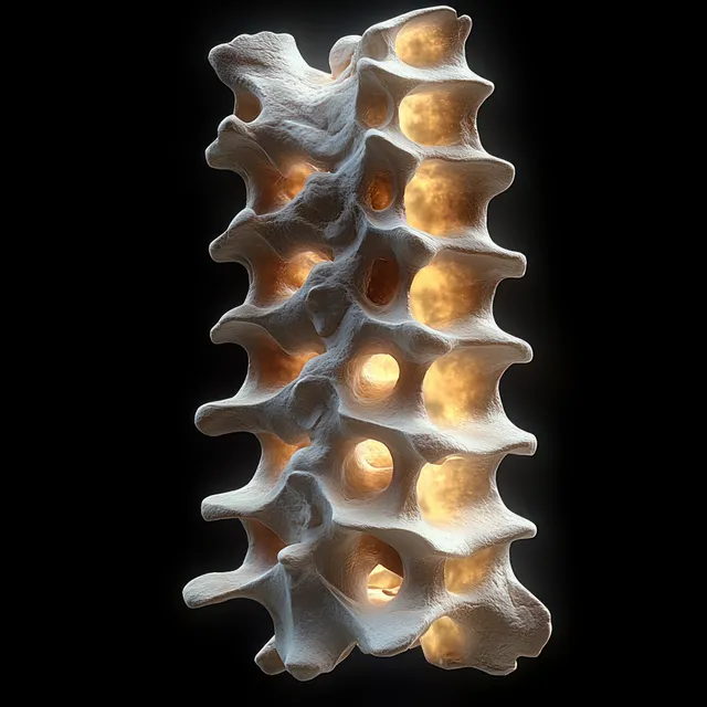
3D vertebral column model with illuminated spinal discs isolated on a dark background.
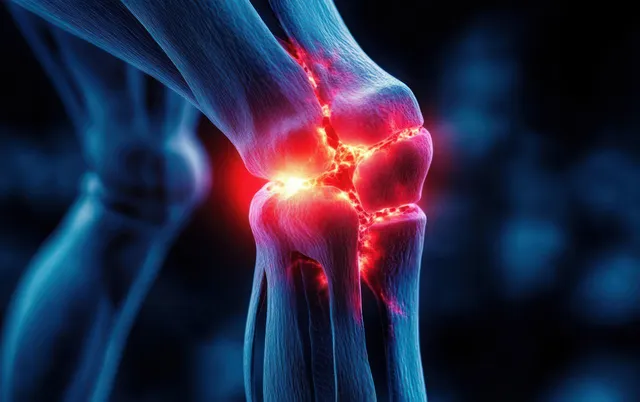
Digital knee joint model with glowing red inflamed areas around the joint and surrounding bones.
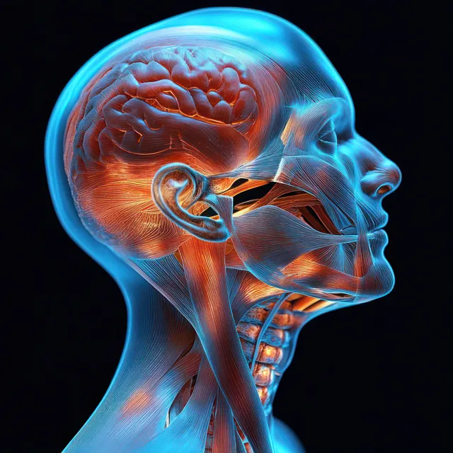
Profile view anatomical illustration of the human neck with highlighted muscles, nerves, and connective tissues.
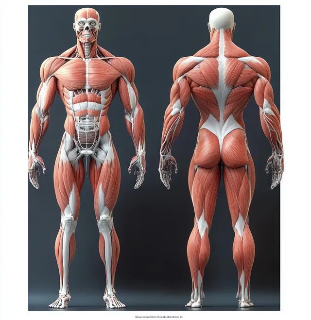
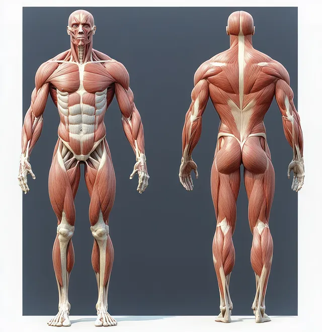
Front and back anatomical illustration of a male figure showing muscles and skeletal bones under rim lighting.
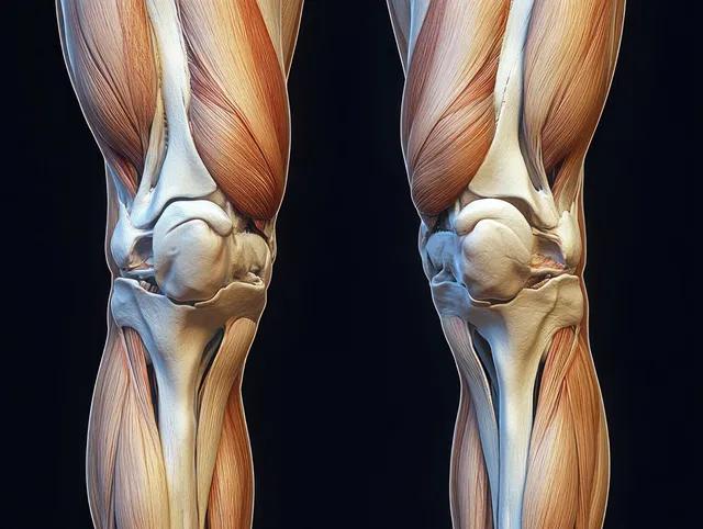
Detailed illustration of human knees showing cartilage, meniscus, ligaments, and surrounding muscles in two mirrored views.
Two-view anatomical illustration of a male body showing muscles, skeleton, and internal organs with dramatic lighting on a dark background.
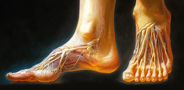
Illustrated feet split into exterior view and cross-section, showing bones and tendons under dramatic dark-to-light lighting.
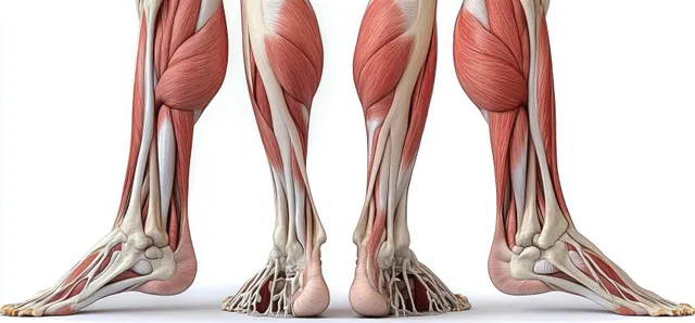
An anatomical cutaway illustration shows leg muscles and bones from front and back views on a white background.
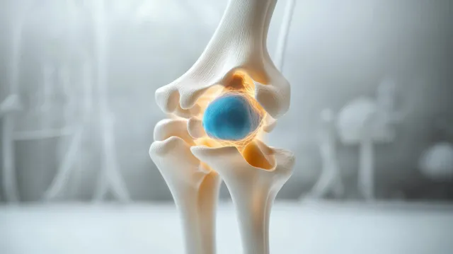
3D anatomical knee joint model with highlighted blue meniscus and cartilage on a softly blurred background.
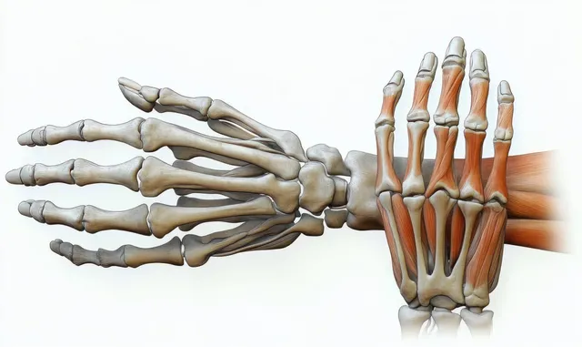
An illustrated human hand cutaway displays the skeleton, muscles, tendons, and layered skin on a neutral background.